
20-year-old female with a 4-year history of right calf pain 12-24 hours after physical activity. A CT scan was done with a “relaxed” phase with the patient lying on the table with the legs in a naturally straight position, and a “flexed” phase with the patient’s toes pointing toward head as far as possible. The relaxed phase showed normal anatomy while the flexed phase showed complete occlusion of the popliteal artery. The Stanford 3DQ Lab technologists provides VR images and CPR Loop movies to show both relaxed and flexed phases in order for the physicians to evaluate the pathology in different body positions, helping the doctors with diagnosis and treatment planning.
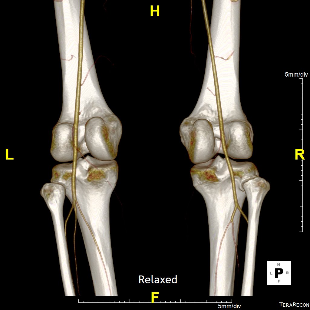
VR image of relaxed phase

VR image of flexed phase
Flexed CPR movie
Relaxed CPR movie

Rhea Liang
3DQ Technologist