Jack Szostak studies the origin and early evolution of life through efforts to design and synthesize a self-replicating protocell capable of Darwinian evolution.
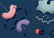
We are interested in the chemical and physical processes that facilitated the transition from chemical evolution to biological evolution on the early earth. As a way of exploring these processes, our laboratory is trying to build a synthetic cellular system that undergoes Darwinian evolution. Our view of what such a chemical system would look like centers on a model of a primitive cell, or protocell, that consists of two main components: a self-replicating genetic polymer and a self-replicating membrane boundary (Figure 1). The job of the genetic polymer is to carry information in a way that allows for both replication and variation, so that new sequences that encode useful functions can be inherited and can further evolve. The role of the protocell membrane is to keep these informational polymers localized, so that the functions they encode lead to an advantage in terms of their own replication or survival. Such a system should, given time and the right environment, begin to evolve in a Darwinian fashion, potentially leading to the spontaneous emergence of novel genomically encoded catalysts and structural molecules.
Figure 1: Protocell model.
Conceptual model of a heterotrophic protocell. Growth of the protocell membrane results from the incorporation of environmentally supplied amphiphiles, whereas division may be driven by intrinsic or extrinsic physical forces. Externally supplied, activated nucleotides permeate across the protocell membrane and act as substrates for the nonenzymatic copying of internal templates. Complete template replication followed by random segregation of the replicated genetic material leads to the formation of daughter protocells.
Image by Janet Iwasa. From Mansy, S.S., Schrum, J.P., Krishnamurthy, M., Tobé, S., Treco, D., and Szostak, J.W. 2008. Nature 454:122–125. Reprinted by permission from Macmillan Publishers Ltd.
Figure 2: Schematic diagram of the growth and division cycle for a multilamellar vesicle.
Figure 3: Overlay of x-ray crystal structures of a native RNA duplex (red) and the same duplex with a single 2′-5′ linkage on each strand (blue). The sequence of each RNA strand is 5′-CCGGCGCCGG-3′, and the single 2′-5′ linkage is between the central C and G residues in the blue structure. The two structures have a similar overall geometry, with local distortions confined to the vicinity of the 2′-5′ linkage.
Left: A vesicle consisting of a fatty acid membrane (red and gray structure) contains RNA primer template complexes (blue and gray). Complexes of Mg2+ and citrate are present inside and outside of the vesicle.
Middle: Activated nucleotides are added to the outside of the vesicle and diffuse across the membrane to the internal space.
Right: The activated nucleotides extend the primer (red) by copying the RNA template.
Adapted from Adamala, K., and Szostak, J.W. 2013. Science 342:1098–1100.
We hope that our explorations of the chemistry and physics behind the emergence of Darwinian evolution will lead to explanations for some of the universal properties of modern cells, as well as explanations of how modern cells arose from their simpler ancestors. As we explore these fundamental questions we are also on the lookout for chemical or physical phenomena that might have practical utility in biomedical research.
Movie 1: An oleate vesicle grows over a period of ~25 minutes after the addition of 5 equivalents of oleate micelles, and divides under the influence of mild fluid agitation.
The first cells would have required a cell membrane that was very different from the membranes of modern cells, which rely on sophisticated protein channels and pumps to control the influx and efflux of small molecules. Since primitive cells lacked highly evolved protein machinery, their membranes would have had to allow nutrients to diffuse into the cell spontaneously. Similarly, membrane growth and division would have had to proceed in the absence of sophisticated biological machinery. How might this have been possible? We have found that membrane vesicles built from simple amphiphilic molecules, such as fatty acids, are excellent models for the compartment boundaries of primitive cells because of their high permeability to polar molecules, such as nucleotides, and because of their dynamic properties, which allow for surprising growth mechanisms.
We have recently discovered several prebiotically realistic processes that lead to the coupled growth and division of fatty acid vesicles. In the simplest process, the addition of fatty acid micelles to preformed vesicles causes the vesicles to grow into long, fragile filamentous structures (Figure 2; Movie 1). These readily divide into multiple smaller daughter vesicles in response to gentle agitation, such as might result from waves on a pond. We are continuing to study this pathway to obtain a better understanding of the underlying physical processes. More recently, we have found that simple concentration by evaporation can drive the growth of vesicles, as free fatty acids in solution partition into the membrane during progressive concentration.
Vesicle growth can also occur as a result of competition between vesicles for limiting fatty acids. We first observed competitive growth when we mixed osmotically swollen vesicles with empty vesicles; after mixing, swollen vesicles grow larger by absorbing fatty acids from the relaxed vesicles, which shrink. More recently we have observed similar phenomena following the mixing of plain fatty acid vesicles with vesicles that contain a small fraction of either phospholipids or hydrophobic peptides. In this case, the vesicles that contain phospholipids (or hydrophobic peptides) grow at the expense of the plain fatty acid vesicles, which shrink. This exciting result implies that any heritable catalyst, such as a ribozyme that could synthesize phospholipids or hydrophobic peptides, would confer a strong selective advantage on its host protocell, potentially leading to the beginning of Darwinian evolution at the cellular level.
Concurrently with our work on vesicle replication, we are addressing the other major challenge in the synthesis of a protocell—the development of a self-replicating genetic polymer. Despite many years of effort, complete cycles of chemical (i.e., nonenzymatic) replication of RNA have not been achieved. Much of our current effort is therefore focused on solving the problems that have blocked previous efforts to replicate RNA without enzymes. This chemical approach to genomic replication broadens the possibilities for the first genomically encoded catalysts, because they would not necessarily have been involved in replication but could have played a role in other processes, such as metabolism.
To better understand the process of nonenzymatic RNA copying, we are intensively studying the binding of activated nucleotides to template strands and the subsequent chemical reaction that results in primer extension through the sequential addition of monomers. We are using nuclear magnetic resonance (NMR) methods and x-ray crystallography to study monomer binding, and studies of reaction kinetics together with molecular modeling to investigate the reaction mechanism. We expect that an improved understanding of the reaction will aid our efforts to improve the rate and fidelity of the RNA-copying process.
One aspect of nonenzymatic RNA copying that was long thought to be a serious problem is that chemically assembled RNA strands have a heterogeneous backbone, with 2′-5′ linkages mixed in with the "correct" 3′-5′ linkages. However, our studies show that the RNA backbone is surprisingly flexible and is able to accommodate 2′-5′ linkages with minimal distortion (Figure 3). As a result, it is still possible to form functional RNAs, such as aptamers and ribozymes, from RNA that contains a high level of 2′-5′ linkages. Remarkably, such linkages have a beneficial effect, in that they lower the melting temperature of the RNA duplex, allowing post-copying strand separation to occur by heating. As a result of these studies we now think that 2′-5′ linkages, far from being a problem with RNA, may in fact be one of the reasons that RNA emerged as the first biopolymer.
The integration of replicating RNA with a replicating compartment boundary system provides numerous opportunities for both positive and negative interactions. Clearly the two subsystems must be compatible for the protocell as a whole to reproduce and evolve. For many years there appeared to be a fundamental incompatibility between RNA replication, which requires high levels of Mg2+ cations, and fatty acid membranes, which are destroyed by moderate levels of Mg2+. However, we have recently found that when Mg2+ ions are bound to citrate, membranes are protected but RNA copying can still proceed. This has allowed us to carry out RNA-copying reactions within fatty acid vesicles, simply by adding activated nucleotides to the outside of vesicles that contain encapsulated primer-template complexes (Figure 4). In addition to being a major step toward the synthesis of a complete protocell, this experiment is significant because it implies that early protocells could have been heterotrophs that grew by taking up nutrients that were synthesized in the external environment.
Our current work is aimed at extending our protocell model by demonstrating complete cycles of template replication within replicating vesicles. If we are able to reach that goal, we hope to observe the spontaneous evolution of adaptive innovations in this relatively simple chemical system. The nature of such adaptations may provide clues as to how modern cells evolved from their earliest ancestors. Ultimately this research may also tell us whether the conserved biochemistry of life is driven by chemical necessity, or whether biochemically very different forms of life are also possible.
This work was also supported in part by grants from the National Science Foundation, NASA, and the Simons Collaboration on the Origins of Life.