ICMIC@Stanford 2010 - 2015
In this renewal grant (2010 - 2015) our overall vision now emphasizes the application and extension of molecular imaging to translational research and clinical applications. Certainly, we also want to continue to i) exploit molecular imaging in the extraction of basic information from animal models and from pre-clinical studies ii) provide new information on tumor diagnosis, initiation, progression, and responses to therapy in these models and iii) develop new imaging technologies. We anticipate that our selection of Developmental Fund projects in the future (for years 2-5 of the renewal) will include funding proposals from outstanding basic science cancer researchers who will want to integrate molecular imaging techniques into their programs. However, our major goal for the ICMIC@Stanford in its second five-year cycle is to provide the groundwork, particularly in the Research Component projects, to integrate molecular imaging into translational studies that move into clinical cancer applications. Therefore, three of the four Research Component projects (RPs 1-3) and one of our Developmental Fund projects (DP2, Willmann) will promote the interactions of basic cancer/molecular imaging researchers with clinical researchers in the translation of studies from animal models on cancer initiation, progression, diagnosis, staging, and response to therapy - to clinical application. We specifically bring together numerous physicians in RPs 1-3 with molecular imaging scientists in order to further bridge our clinical and scientific community. This will allow us to continue to accomplish our long-term vision of translating molecular imaging strategies into the clinic. We also form important scientific links to our NCI Funded CCNE U54 (RP1,3) and NTR U54 (RP2) programs through use of in vitro nanosensors and intraoperative microscopy, respectively. This will help to further accelerate our ability to bring important state-of-the-art solutions to cancer research and cancer patient care.
The Molecular Imaging Program at Stanford is directed by Dr. Sanjiv Sam Gambhir, Professor of Radiology and Chair of the Department of Radiology, and is co-directed by Dr. Chris Contag, Professor of Pediatrics and of Microbiology and Immunology and, by courtesy, of Radiology and Director of Stanford Center for Innovation in in vivo Imaging (SCI3). Together, Drs. Gambhir and Contag form a highly unique leadership team that spans the diversity of disciplines involved in the field of multimodality molecular imaging. The MIPS currently has 13 full members (including Drs. Contag and Gambhir) from 5 different departments and 30 associate members from 11 different departments. Full members have greater than 70% of their research efforts focused on multimodality molecular imaging whereas Associate members are actively using multimodality molecular imaging but have a focus in another scientific discipline.
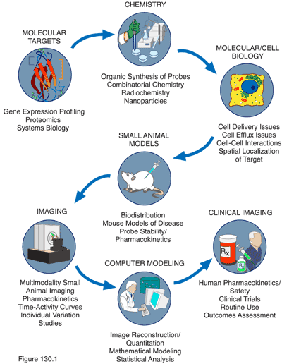
Multimodality Molecular Imaging requires the interactions of several disciplines as shown below. The MIPS covers the breadth of disciplines needed to perform successful research in multimodality molecular imaging. It is important to note that although many of the MIPS faculty are focused on cancer research, several are studying other areas including neurosciences and cardiovascular biology.
Our mission is to establish, for the long term, a multimodality in vivo cellular and molecular imaging program to study neoplastic disease and better link pre-clinical models with clinical cancer management through advances in multimodality molecular imaging. The program will address key issues in cancer research from the level of oncogenesis to using imaging for optimizing anti-cancer therapies in pre-clinical models and in cancer patients. The goals of this program are to:
- Maximize interaction among multidisciplinary investigators for a coordinated effort in molecular and cellular imaging of cancer
- Coordinate and manage the multidisciplinary programmatic cores and collaborative research efforts, and integrate these into the specialized research activities of the University for an effective research endeavor that pushes the envelope of scientific discovery
- Understand basic biological mechanisms of cancer that will lead to intervention strategies that will aid in the design and testing of innovative therapies that strike at the molecular foundation of oncogenesis and the minimal disease states that lead to relapse
- Maximize the use of the specialized resources and program strengths with integrated projects that aim to advance novel imaging approaches in oncology
- Provide training opportunities for students and scientists at all career levels to advance the field of in vivo cellular and molecular imaging, nationally and internationally.
Overview of ICMIC Structure

The above figure is an organization chart with names of all members of the ICMIC@Stanford including members of the Executive Committee, our Administrative Assistant, the Internal Advisory Board, External Advisory Board, Directors of Research Components, Specialized Resources, Developmental Projects and make-up of the Career Development Program. There are 18 faculty members in 10 different disciplines involved in the ICMIC@Stanford. There are also 35 additional MIPS members, many of whom already function as mentors to our trainees.
