Stanford Moyamoya Center
MOYAMOYA BANNER HERE
The Vascular Neurosurgery program at Stanford provides state-of-the-art care and management of all forms of cerebrovascular disease. Stanford is a quaternary referral center for complex neurovascular lesions. The Vascular Neurosurgery team collaborates closely with neuroradiologists and stroke neurologists to plan treatments for challenging cerebrovascular diseases using endovascular embolization, radiosurgery, arterial bypass grafting, and microsurgery.
What is Moyamoya?
Moyamoya disease is a progressive disorder that affects the blood supply to the brain. It is rare, but is being detected more frequently with recent advances in diagnostic imaging.
It can occur at any time, but is most commonly diagnosed in childhood between ages 5 - 15 and during adulthood between ages 30 - 40.
History of Moyamoya
Moyamoya was first identified by the Japanese, who named it for the appearance of a “puff of smoke” that appeared on the cerebral angiogram. It is more common in Asians, but can affect any population.
Moyamoya disease is characterized by a progressive narrowing of the internal carotid arteries leading into the brain, and usually affects both sides of the brain.
The cause of arterial narrowing is unknown, but does not appear related to atherosclerosis or inflammation of the arteries.
As the vessels narrow the brain receives less blood. This can result in temporary symptoms such as:
- Headaches
- Numbness or weakness in the extremities
- Difficulty speaking
- Stroke
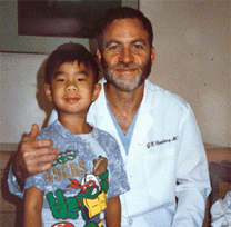
|
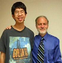
|
| Dr. Steinberg with Moyamoya patient 1993 |
Dr. Steinberg with the same Moyamoya patient 2004
|
Symptoms
Children typically have symptoms including:
- Strokes (sustained weakness or numbness in an arm or leg, difficulty speaking, visual abnormalities or problems walking)
- Transient ischemic attacks, or TIA’s (temporary stroke-like symptoms that don’t last long)
- Headaches
- Progressive cognitive or learning impairments
Children also often experience temporary weakness in one or more of their extremities during strenuous physical activity or when crying. Adults can also present with brain hemorrhage causing neurologic symptoms in addition to nonhemorrhagic strokes, TIA’s and headaches.
Moyamoya sometimes occurs along with other disorders such as Down’s Syndrome, brain AVM’s (arteriovenous malformations), and neurofibromatosis. We have also seen it in patients who had previous brain radiation for tumors.
Diagnosis
Based on the patients’ symptoms and history, the physician may order one or all of these tests before making a decision about treatment.
Magnetic Resonance Imaging (MRI)
This allows physicians to examine brain structures and detect any strokes that might have occurred.
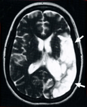 |
The arrows on this MRI of the brain shows a patient with a large stroke from moyamoya. |
Cerebral Angiogram
This is the definitive test that confirms the diagnosis of moyamoya. “Contrast” (or dye) is injected into arteries to reveal the anatomy of the arteries of the brain and scalp. This is a minimally invasive test that requires patients to stay at the hospital for several hours. This test assesses the severity of moyamoya and its results guide treatment options, which are determined by how severe the disease is and what the external (scalp) blood supply is.
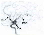 |
Slide view of a normal angiogram.
MCA=middle cerebral artery
ACA=anterior cerebral artery
ICA=internal carotid artery |
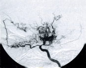 |
Angiogram of a patient with moyamoya disease. You can see the MCA and ACA vessels are imparied and moyamoya vessels have appeared along with other collateral vessels. |
Xenon CT Scans
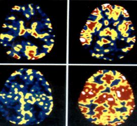
This test reveals blood flow to regions of the brain to determine if enough blood is reaching all areas. Patients breathe xenon (an odorless, colorless gas), which acts as a contrast agent to show regions of low and high blood flow.
Pre-Operative
Post Operative
|
Pre-Diamox
Post-Diamox |
Note the areas of red, which indicate a higher volume of blood flow to the brain when challenged with Diamox.


