2015 MIPS News
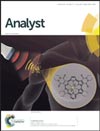
MIPS Research Featured on the Inside Cover of AnalystMay 18, 2015 As discussed in the paper entitled, "Parts per billion detection of uranium with a porphyrinoid-containing nanoparticle and in vivo photoacoustic imaging," by I-Ting Ho, PhD, et al., researchers have developed an in vivo imaging method to quantitate uranium. This approach uses a porphyrin-based molecular probe that is only aromatic—and thus optically active—in the presence of a uranyl cation. This approach may eventually have defense applications. |
Dr's Witney and James Received SNM Alavi Mandell AwardApril 15, 2015 Dr. Tim Witney and Dr. Michelle James each received the Alavi Mandell Award from the Society of Nuclear Medicine for their respective publications: "Preclinical Evaluation of 18F-Fluoro-Pivalic Acid as a Novel Imaging Agent for Tumor Detection" The Alavi-Mandell Award recognizes outstanding contributions to the field from young scientists, fellows, or physician residents who have published (as first authors) excellent original articles in the Journal of Nuclear Medicine. In 2015, SNM only gave out 4 Alavi Mandell Awards. Congratulations to Dr. Witney and Dr. James! |
Aaron Mayer and Surya Murty Awarded NSF FellowshipsMarch 31, 2015 Please join us in congratulating Aaron Mayer and Surya Murty who received 2015 NSF Graduate Research Fellowships. The NSF Fellowship supports individuals early in their graduate careers that have demonstrated potential for significant achievements in science and engineering. Aaron is a first year bioengineering PhD student whose NSF proposal include plans for the development of a molecular imaging toolkit to help better understand, monitor, and improve cancer immunotherapies. Surya is also a first year bioengineering PhD student and this fellowship will fund three years of his degree where he will investigate imaging and improving immunotherapies against brain tumors. Congratulations, Aaron and Surya! |
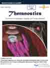
MIPS Research Featured on the Cover of TheranosticsMarch 30, 2015 The paper entitled "Theranostic Mesoporous Silica Nanoparticles Biodegrade after Pro-Survival Drug Delivery and Ultrasound/Magnetic Resonance Imaging of Stem Cells," by Paul Kempen, PhD, et al. from the Multimodality Molecular Imaging Lab was highlighted on the cover of the journal Theranostics. |
Dr. Daldrup-Link Elected to the American Society for Clinical Investigation (ASCI)March 18, 2015 Please join us in congratulating Dr. Heike Daldrup-Link in her election to the American Society for Clinical Investigation (ASCI)! She was nominated by ASCI members Dr. Sanjiv Sam Gambhir and Dr. Linda Boxer for her contributions in the field of translational cellular imaging. Dr. Daldrup-Link introduced tumor characterizations via the enhanced permeability and retention effect and lead a first multi-center clinical trial on nanoparticle-enhanced MR imaging of breast cancer to prove this concept in patients. She discovered a new approach for MR imaging of tumor associated macrophages (TAM) with iron oxide nanoparticles in mouse models and obtained an IND to carry out the first TAM imaging trial in pediatric patients. This approach will be used in the future to monitor new anti-CD47 mAb immunotherapies in patients. In collaboration with Dr. Rao, Dr. Daldrup-Link developed tumor-enzyme activatable theranostic nanoparticles, without side effects for combined cancer imaging and therapy. She recently introduced a novel radiation-free whole body staging test for children with cancer (Lancet Oncology 2014). Dr. Daldrup-Link's cellular imaging studies also yielded several new and patented ideas for in vivo imaging of stem cell transplants, establishing immediately clinically applicable technologies for: in vivo stem cell tracking with FDA-approved nanoparticles, in vivo imaging of stem cell rejection processes with immune-cell targeted tracers, and MRI-detection of stem cell apoptosis with a caspase-activatable contrast agent. These cellular imaging tools provide a truly new way to evaluate stem cell physiology beyond simple cell detection, expanding our understanding of in vivo stem cell engraftment outcomes. The ASCI is one of the nation's oldest and most respected medical honor societies of physician-scientists, those who translate findings in the laboratory to the advancement of clinical practice. Founded in 1908, the Society is home to more than 3,000 physician-scientists from all medical specialties elected to the Society for their outstanding records of scholarly achievement in biomedical research. The ASCI represents active physician-scientists who are at the bedside, at the research bench, and at the blackboard. Many of its senior members are widely recognized leaders in academic medicine. The ASCI is dedicated to the advancement of research that extends our understanding and improves the treatment of human diseases, and members are committed to mentoring future generations of physician-scientists. The ASCI considers the nominations of several hundred physician-scientists submitted from among its members each year and elects up to 80 new members each year for their significant research accomplishments. Because members must be 50 years of age or younger at the time of their election, membership reflects accomplishments by its members relatively early in their careers. Congratulations, Dr. Daldrup-Link! |
MIPS Research Uses Tumor-activatable Minicircles for Early Detection of CancerMarch 2, 2015 New work from the Gambhir Lab published in PNAS uses a unique strategy to force tumor cells (if they exist) to produce a blood biomarker that would otherwise not be present. This approach holds significant promise as a new way to tackle the early detection of cancer because it is not dependent on molecules that cancer cells naturally shed that enter the blood. Read the abstract
|
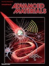
MIPS Research Featured as the Inside Cover in the Advanced MaterialsFebruary 4, 2015 The paper entitled "Perylene-Diimide-Based Nanoparticles as Highly Efficient Photoacoustic Agents for Deep Brain Tumor Imaging in Living Mice," by Quli Fan, PhD, et al. from the Cancer Molecular Imaging Chemistry Lab was highlighted by Advanced Materials as the Inside Cover. |


