


Bacteria are everywhere: floating through air, drifting in water, clinging to surfaces. So it’s not surprising that many animals and plants have formed beneficial relationships with the microorganisms.
We are surrounded by examples of bacteria helping animals and plants. Bacteria can protect plants against extreme drought. The bacterial mix living in an animal’s gut makes digestion possible. The good bacteria that coat skin fight off harmful microbes. But as scientists delve deeper into the ties that bind animals, plants, and bacteria, they’re finding some unexpected, fundamental connections: in some cases, the chemical signals released by bacteria trigger development. They may even hold clues as to why organisms became multicellular.
Two recent studies reveal the molecular nature of some of these chemical cues. But what do the bacteria get out of the relationships? “Animals evolved in a bacterial world,” says Dianne Newman, an HHMI investigator who studies bacteria at the California Institute of Technology. “I think we’re just at the very beginning of really starting to appreciate that and what it means.”
Bacterial Welcome Mat
The tubeworm Hydroides elegans begins its life as a tiny larva, floating freely through the ocean. Eventually, it needs to grow up, settle down on a hard surface such as a rock or the hull of a boat, and build its “house”—a calcified, tube-like outer shell. The trigger for this transition from free-swimming larva to stationary juvenile comes from a carpet of bacteria called a biofilm. The biofilm covers the surface where the H. elegans lands to settle down. The transformation is fascinating, but it’s also a bane for the shipping industry. This accumulation of bacteria and marine animals, known as biofouling, creates extra drag on boats and increases fuel consumption.

Michael Hadfield, a biologist at the University of Hawaii, has spent 25 years studying the tubeworm as it settles into adulthood. He’s focused on one of several biofilm-forming bacteria that trigger the metamorphosis—Pseudoalteromonas luteoviolacea, or P. luteo, which is by far the most efficient. In 2012, Hadfield’s graduate student Ying Huang zeroed in on four genes in the bacteria that trigger tubeworm transformation. But the specific cue that sparked the worm’s lifestyle change remained unknown.
Two years later, Nicholas Shikuma, a postdoctoral fellow in Newman’s lab, collaborated with Hadfield to uncover the products of the four genes. “I thought the bacteria were putting out a peptide or something that the larvae would have specific receptors for,” says Hadfield. It turns out the tubeworms were responding to something much bigger and more complex: a structure composed of hundreds of protein components. “This really changed our whole perspective,” says Hadfield.
Big MACs
Shikuma discovered that the signals produced by P. luteo were actually needle-like macromolecules that he and his colleagues dubbed metamorphosis-associated contractile structures, or MACs. Each MAC consists of three parts: a narrow tube that can be used as a projectile, a hollow sheath that surrounds the tube and pulls back when triggered, and an anchoring baseplate. The structures bear a strong resemblance to the syringe-like tails found in some bacteriophages—a type of virus that infects bacteria (the viruses use the narrow tube contained in the needles to pierce bacterial envelopes and then inject a payload of viral DNA inside).
“The MACs are like spring-loaded molecular daggers,” explains HHMI Investigator Grant Jensen, who collaborated with Shikuma to get an up-close look inside the bacteria by using a technique called electron cryotomography. “Their outer sheath contracts like an accordion, ejecting the inner rod and its chemical payload into the target.”
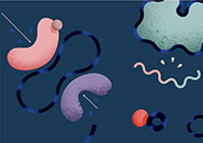
Even more intriguing was the finding that the needles formed porcupine-like arrays that, when fully extended, were larger than the cells that produced them. At the base of the array, the MACs are tightly gathered like the stems in a bouquet of flowers. At the other end, the rods project out like a bundle of tiny spring-loaded syringes.
, that are bound together in porcupine-like arrays. Illustration courtesy of Grant Jensen.")
“This type of assemblage was structurally really different from what had previously been seen for phage tail-like particles,” says Newman. “And what’s more, unlike previous cases where such particles were linked to pathogenesis, the MACs mediate a beneficial interaction.” The group published its findings in the January 31, 2014, issue of Science.
Only about 2 percent of P. luteo cells in a given biofilm produce MACs, and they give themselves fully to the process. They convert their entire contents to MACs, packing their insides wall to wall with the arrays. Eventually, they get so full they burst, releasing the arrays, which spring open like umbrellas.
Although the scientists aren’t exactly sure how the MACs trigger metamorphosis, they think that the needles may act like molecular guns, firing a payload of who knows what into the H. elegans larva that starts the change from innocuous organism to invasive pest.
Alternatively, perhaps the tubes themselves act as arrows to puncture the larva’s cells. “To me they look like a whole bunch of cocked crossbows,” says Hadfield. “If they hit the right cell—a sensory cell on Hydroides—and poke holes in it, it could be sufficient to trigger metamorphosis.”
How the bacteria benefit from this relationship and why they produce MACs are also unclear. Perhaps the worms are protecting P. luteo by ingesting their zooplankton predators. Whatever the mechanism and the relationship, it’s clear that the bacteria are signaling to the tubeworms that it’s time to grow up.
A Quest for Colonies
Much like H. elegans, the tiny water-borne organisms called choanoflagellates rely on bacterial cues to enter a new stage of life. Choanoflagellates spend their time gorging on bacteria in oceans, lakes, ponds, and even puddles. Generally, they are single celled. But some species, like Salpingoeca rosetta, can also form large colonies. As its name implies, as S. rosetta cells divide, they arrange themselves into a rosette, radiating around a central point. Little is known about the lifestyle of these organisms, but since they are among the closest living relatives of all animals, their biology may shed light on how and why our ancestors became multicellular.

HHMI Investigator Nicole King became fascinated by choanoflagellates when she was a postdoctoral fellow in 2000. Since then, she has been on a quest to figure out how and why they form colonies. In the wild, S. rosetta readily develops into rosette colonies. In the lab, it was a different story. For years, King managed to coax the organisms to develop into colonies only once in a while.
So she decided to sequence the genome of S. rosetta, hoping for clues to this developmental process. That’s when undergraduate researcher Richard Zuzow made a serendipitous discovery. To prepare the cells for sequencing, Zuzow needed to remove contaminating bacteria. So he treated the cells with antibiotics. Certain cocktails of antibiotics, he noticed, caused the choanoflagellates to stick together, while others prevented rosette formation.
“Even when we washed out the antibiotics, the colonies never came back,” explains Rosie Alegado, who at the time was a postdoc in King’s University of California, Berkeley lab. “Either the antibiotics were directly affecting choanoflagellates or we killed off something in the culture that was triggering this effect.”
A Simple Signal
It turned out to be the latter. Zuzow figured out that certain bacteria—Algoriphagus machipongonensis—were prompting rosette formation; the antibiotics used to prep the cells for sequencing were killing off the bugs. Alegado, now at the University of Hawaii, teamed up with Jon Clardy, a natural products chemist at Harvard Medical School, to purify the substance that was causing the rosettes to form.
It was a much simpler signal than the MAC arrays that spur tubeworm metamorphosis. A. machipongonensis releases a lipid molecule that belongs to the sulfonolipid family. Similar compounds had been seen before, but their functions were unknown. The team named the molecule rosette-inducing factor 1, or RIF-1, and published its findings in eLife on October 15, 2012.
“First we find that a bacterium is actually regulating whether choanoflagellates are single celled or colonial. That’s exciting because choanoflagellates eat bacteria, and they’re getting cues about their environment from the bacteria,” King says. “Then we find out that it’s the special class of molecules that hasn’t been characterized before.”
As with the tubeworms, many questions about the choanoflagellate-bacteria relationship remain. Alegado and King think that RIF-1 is released into the water via vesicles that “bleb off” the bacterial membrane. These lipid bubbles either fuse with hydrophobic molecules in S. rosetta’s membrane, or they are engulfed by the choanoflagellate. Then they trigger rosette formation—and this is where things get murky.
“At this point, I feel very comfortable talking about how things are happening,” says King. “I’m just less certain about why.” For example, she has no idea why A. machipongonensis produces RIF-1 or why S. rosetta responds to it. One hypothesis for the latter is that colonies are better at capturing bacteria, so the choanoflagellates form rosettes to better exploit a resource.
Both the tubeworm and choanoflagellate studies illustrate that, in some cases, intricate relationships have evolved between bacteria and the organisms they live with. They also raise many questions about how and why the relationships formed, and whether or not they are exceptions or the norm. “They are both anecdotes of how bacteria are intentionally or unintentionally driving the behavior of multicellular and transiently multicellular organisms,” says Alegado. “The two papers indicate that there’s a rich chemical dialogue going on that we know very little about. It just shows that we have to continue to look.”


