


Bi-Chang Chen spent the fifth day of his postdoctoral fellowship in a rented van headed for Cape Cod. He’d barely had time to unpack at Janelia Farm before he was enlisted to help take apart the very microscope he had come to Virginia to work on. Its lenses, mirrors, lasers, and other components were now fastidiously packed and stowed in the back of the van, traveling north on I-95. Chen was about to undergo what his new boss, Janelia Farm group leader Eric Betzig, was calling “trial by fire.”
Chen and his new colleague, Liang Gao, were transporting the Betzig lab’s newest technology to the Marine Biological Laboratory (MBL) in Woods Hole, Massachusetts—a seaside hub of scientific activity with an educational program that draws hundreds of students and faculty from around the world each summer. It would take a full day to reassemble its components into what the Betzig group calls a Bessel beam plane illumination microscope—a high-speed, high-resolution, three-dimensional imaging technology that gives extraordinarily detailed views of cellular processes in action.
The plan was for Gao and Thomas Planchon—developers of the new microscope—to guide MBL students and faculty through the imaging process as they examined their specimens. Chen would have to learn quickly how to operate the microscope, because the team expected a steady stream of users, all demanding expert attention. In exchange, the Betzig team hoped to learn exactly what biologists need their microscope to do.

The Bessel beam plane illumination microscope, or “Bessel sheet,” was a work in progress. Betzig’s team had designed it to answer an obvious need of biologists—the ability to visually track movement of the tiniest structures inside living cells. In March 2011, they had described the new technology online in Nature Methods, including as evidence of its power startlingly vivid portrayals of chromosomes sorting themselves in preparation for cell division and a lively cell surface sending out long, thin projections that quickly retract and reappear elsewhere.
The short movies had generated considerable excitement among biologists: there was no doubt that the new microscope surpassed existing technologies in rapidly creating high-resolution, three-dimensional images without damaging living cells. But for Betzig’s team, this acclaim was not the ultimate goal. “We’re not developing technology for technology’s sake,” he says. “We want to create technologies that will have a broad and deep impact on biomedical research.”
Maximizing the microscope’s impact had been foremost in Betzig’s mind from its earliest development. A physicist and engineer by training, Betzig listens carefully to biologists, and their top priorities have not changed since he began developing imaging technologies more than 30 years ago. Biologists want to see smaller structures. They want to see them with more detail, with more speed, and deeper inside a tissue. And they want to see them in healthy, living cells that are unperturbed by the tool they are using.
|
WEB EXTRA |
|
 |
Working Retreat |
Betzig and his team had created the Bessel sheet microscope to answer those demands, and after three years of development, they were convinced the instrument could offer researchers unprecedented views of dynamic subcellular processes. But many of the biologists at Janelia Farm are neuroscientists interested in understanding how neurons work together in complete circuits in the brains of fruit flies or mice, and, Betzig says, Bessel beam technology is not ideal for the size and thickness of the samples they want to image. “Our mandate is to develop new imaging technologies for biology, not necessarily just for systems neuroscience. So if we want to find users for these techniques, we have to look outside.”
Word was beginning to spread about the power of the Bessel sheet, but the cultural divide between microscopists and biologists still frustrated the Betzig team. “It takes a surprisingly long time to get new microscopes to biologists,” Gao says. “So we decided to take our latest microscope to Woods Hole to introduce it to them.”
Rapid-Fire Days
The Marine Biological Laboratory is situated on a narrow, salt-swept piece of land in the far southwest corner of Cape Cod. Tourists unload bikes at the nearby ferry landing; fried clams, homemade ice cream, and other indulgences of a beach vacation are in ready supply. The tiny village of Woods Hole wakes up earlier than the usual beach town, however, and working scientists vastly outnumber vacationers.
Hundreds of scientists flock to Woods Hole in summer to immerse themselves in MBL’s unique culture for a few weeks or months. Distanced from routine distractions, most visitors find the MBL experience gratifyingly intense. Between coursework, research, and discussions and debates, time in the lab routinely stretches until 2:00 a.m., for students and faculty alike. “We’re not there to go to the beach,” Gao stresses. “There’s a vacationy feel here; it’s nice outside,” says Betzig, who set aside two weeks during June and July to work with Bessel sheet users at MBL. “But you’re only here a short time, and everyone works really long days.”
Page 2 of 4
So when Chen, Gao, and their microscope parts reached Woods Hole at the end of June, they wasted no time. They were greeted by Jim and Cathy Galbraith, a husband and wife team who have taught in the neurobiology course for seven years, and a small crew ready to help maneuver an 800-pound optical table, on loan from Thorlabs, into a tiny, windowless room in the basement microscope facility. The top of the table went in first, suspended on a scissor lift until its vibration-eliminating supports were positioned underneath. From there, the team followed Gao’s lead. Modular segments of the microscope were unwrapped and bolted to threaded holes in the tabletop. A tower of electronic equipment—the control panel—was erected in the cramped room’s remaining space. Cables were connected; alignment measured; blue, green, violet, and infrared lasers tested.
Gao knew what he was doing: he’d built this version of the microscope—a second-generation version, more compact and portable than the original built by Planchon at Janelia. And last summer, he and Planchon had accompanied the Bessel sheet on its first trip to Woods Hole, when it was so new Betzig was calling it “bleeding-edge.” That visit had been grueling: two weeks of 18-hour days, imaging the very first living cells to be viewed with the microscope, save one quick run-through back at Janelia Farm with the Galbraiths. But the benefits were enormous. During the rapid-fire days, the team tested a panoply of samples and introduced the microscope to a range of users. The Bessel sheet’s early successes at Woods Hole became the vivid movies the team published in its Nature Methods paper, which is coauthored by the Galbraiths, whose own labs are at the National Institutes of Health (NIH). And throughout the following year, the Betzig lab hosted collaborators at Janelia Farm to advance projects initiated during those fervent weeks.
So when Betzig suggested they repeat the experience this past summer, Gao’s first thought was the challenge of transferring the delicate equipment to its temporary home. “It’s almost like starting a new lab,” he says. But he knew it would be worth the long drive and the long days that would follow, particularly if they extended their stay to work with a wider group of biologists.
|
WEB EXTRA |
|
 |
Skin Development in C. elegans |
 |
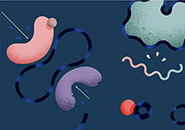
Dividing Chromosomes |
 |
Surface of a HeLa Cell |
They chose a four-week period during which their microscope could be integrated into two courses. Gao and Planchon made plans to helm the microscope in two-week shifts, while Chen would stay the entire four weeks to learn the intricacies of the Bessel sheet and become familiar with the language of its users. Betzig would join the team for the weeks in the middle.
During the first two weeks, the Bessel sheet would help physiology students working with Jennifer Lippincott-Schwartz from the NIH watch cells migrate or break down their unwanted parts; in the second half of the microscope’s stay, neurobiology students working with the Galbraiths could use it to visualize the processes that drive nerve cell function. Spare moments would be tightly scheduled with MBL faculty curious to see what the Bessel sheet might reveal about their own samples.
The Photon Budget
The Bessel sheet was the newest imaging technology at Loeb Laboratory, MBL’s educational hub, but it was not the only one. The basement and first-floor hallways were crowded with bulky crates emptied of sophisticated microscopes on loan for the summer. Course directors had collected commercial instruments for super-resolution imaging (including Zeiss’s ELYRA, based on the concept of photo-activated localization microscopy (PALM), invented by Betzig and Janelia Farm colleague Harald Hess), microscopes designed to peer deep into tissue, and high-resolution methods for three-dimensional imaging of fixed tissue to offer their students new perspectives on biological complexity.
According to Cathy Galbraith, the two-week imaging section of the neurobiology course is designed not just to expose students to cutting-edge technology, but also to help them understand which tools are best suited for particular questions, as well as how the technologies are complementary. Likewise, physiology students experience firsthand how each tool has its strengths—but none can do it all.
Page 3 of 4
It quickly becomes apparent how the Bessel sheet fits into the imaging landscape. Like other fluorescence microscopes, the Bessel sheet detects fluorescently tagged molecules within cells, letting biologists follow specific proteins and watch them interact with one another. The basic technique relies on fluorescent tags that absorb light—typically directed at a sample in a focused laser beam—and rerelease the energy as fluorescence, which the microscope captures to create an image. Up to a point, brighter fluorescence contributes to a clearer image, but there is a trade-off. Too much light exposure can make a cell sick, so living samples don’t survive long under the laser’s glare. What’s more, light destroys fluorophores’ ability to fluoresce, meaning even dead cells can be imaged only a brief time before their signal begins to fade. So, fluorescence microscopy often ends up being an exercise in compromise.
 and Thomas Planchon improve design of their microscopes. Photograph by Jared Leeds")
Capturing fluorescence becomes increasingly crucial as new technologies push the limits of both spatial resolution and speed. Higher-resolution microscopes must extract information faster, more frequently, and from ever-shrinking segments of a sample. Ideally, a microscope would glean information from each photon of fluorescence a sample emits. In practice, however, plenty of photons bounce off an illuminated sample and are lost forever.
Betzig and his team designed their new microscope to take better advantage of what they call the “photon budget.” Most microscopes illuminate a sample from the same objective lens that collects light, directing a beam all the way through the sample even though only a single plane is in focus at a time. At high-resolution, where every photon counts, this is a problem because light in out-of-focus regions activates fluorescence and damages cells, with no imaging payoff.
An alternative is to direct a sheet of light through the side of a sample, via a lens that lies perpendicular to the objective that collects the returned light. This approach confines the excitation much closer to the portion of the sample in focus. It’s an old idea—first proposed more than 100 years ago—that has recently been adapted to high-speed imaging of multicellular organisms. Ernst Stelzer, who was at the European Molecular Biology Laboratory in Heidelberg, Germany, and is now at the Frankfurt Institute for Molecular Life Sciences, resurrected the technique, Betzig says, taking advantage of the reduction in light damage to image living samples repeatedly over prolonged periods.
The thick sheets of light that have made Stelzer’s technique a success, however, obscure the tiny structures inside cells—a problem that Betzig’s team addressed with a Bessel beam. At its core, a Bessel beam is an extremely narrow beam of light that can be swept across a sample to create a thin sheet well suited for imaging subcellular structures. The Bessel’s central beam is surrounded by concentric rings of weaker light, however, which cause the same problems as any unwanted light sources do. So Betzig, Planchon, and Gao devised a few tricks to minimize the contaminating light. Rather than sweeping the beam continuously, they pulse the beam rapidly to eliminate out-of-focus blur—a technique also used in super-resolution microscopy called structured illumination. They combined this pulsing with two-photon microscopy, a method in which weakly exposed regions generate very little fluorescence. Together, these tricks add up to a microscope that collects unusually detailed images without damaging living cells.
3-D Clarity
Gao, Planchon, and Chen help students mount their samples so they can receive the sheet of light created by the Bessel beam at the appropriate angle, then tweak the microscope’s parameters to accommodate variations in thickness, brightness, or background fluorescence. The Bessel beam sweeps quickly through each sample, collecting up to 200 images per second. As the microscope’s computer compiles the two-dimensional images into three-dimensional stacks and the first pictures begin to appear on the monitor, what is astonishing is the extraordinary detail apparent in all three dimensions.
Clare Waterman, director of the MBL physiology course, says she was blown away when she saw the Bessel sheet’s first images of her students’ cells. “I’ve been looking at light microscopy images and 3-D reconstructions for 20 years,” she says. “When you rotate a 3-D reconstruction, you always know what the z-axis is.” That is, even when a microscope produces crisp, detailed images in two dimensions, resolution almost always declines significantly in the third dimension. But the Bessel sheet images were different. “I could not tell what the z-axis was,” Waterman says. The cellular detail was clear from any angle.
The three-dimensional movies of migrating cells, which were grown in a collagen gel so they could move freely in any direction, settled for Waterman what she says had been an unresolved debate in her field. Waterman studies cell motility at the National Heart, Lung, and Blood Institute, and lately, she says, there’s been a big push to find out whether structures thus far observed only in cells squashed flat between a microscope slide and a coverslip are physiologically relevant. But, she acknowledges, “There’s been a reason we’ve been doing stuff on a coverslip for so long: because that’s where optics are good.” Technologies that image in three dimensions lack the spatial resolution to determine, for example, whether migrating cells truly project the long, flat extensions thought to propel their motion. “Well, we could certainly see two-dimensional, flat lamellipods with this [Bessel sheet] system that we could measure the thickness of, in every dimension.”
Page 4 of 4
“It wasn’t like I was pining away for this,” Waterman says. “I had accepted the limits of optical microscopy and I’d learned to interpret my data within those limits.” Once she saw what was possible, she says, “I was doing backflips for a week.”
Watching Embryos Develop

The Bessel sheet gets little rest at Woods Hole. A grid on the wall blocks out time for faculty who want to squeeze into gaps between student projects, but unexpected visitors stream in at all hours. University of California, San Diego, developmental biologist Andrew Chisholm, who visits MBL as faculty in the neurobiology course, says so many people were in and out of the tiny room that the rising temperature sickened the C. elegans roundworm embryos whose developing skin he wanted to image. With the confocal microscopes Chisholm typically uses, he says, “you’re constantly balancing imaging quality with viability of the embryos. With a conventional microscope you really have to settle for images that don’t look very good.” But since the Bessel sheet limits phototoxicity by using far less light, the dying worms were puzzling.
Chisholm and the Betzig team considered other factors, and discovered that if they imaged the developing embryos in the morning—after a few idle hours had let the room cool down—they had better success. Working with Planchon, Chisholm assembled a movie that shows skin cells forming, making junctions with one another, and then forming a sheet that spreads out to encase the growing embryo. Chisholm says the movie, which spans eight hours of early development, is “an order of magnitude better than what we can get away with using the last generation [of imaging technology]. It’s just beautiful.”
User Feedback
By the time the weary team of postdocs disassembles the microscope for the journey back to Virginia, they have imaged developing embryos, cells suspended in growth media, the nervous system of whole worms, cells embedded in collagen, dying cells, and dividing cells. Over the weeks, Chen says, they have taken every opportunity “to talk to biology people, to listen to what they are thinking. Because it’s really, really different from us.”
They have heard users’ frustration about the microscope’s unconventional sample holder, which requires a fussy assembly process to secure the sample in a vertical position—practical from an engineering perspective, but cumbersome for users. They know they need to adapt their optics to handle thicker samples, which introduce aberrations in the imaging process. And there is an unrelenting desire, biologists have told them, for even better spatial resolution, at even faster speeds.
None of these requests are a surprise, the microscopists agree. And they are prepared to address them. The group plans to build a third version of the Bessel sheet, so they can continue to tweak the instrument and test out improvements while the original setups collect data. Chen will begin work immediately to add additional Bessel beams to the microscope, which he says will increase imaging speed while reducing phototoxicity. A new postdoc in the Betzig lab, Kai Wang, will introduce adaptive optics into the system, and research scientist Lin Shao, who introduced the current methods of analyzing the data generated by the Bessel microscope, will continue to improve those algorithms. Thomas Planchon, now an associate professor at Delaware State University, plans to develop similar microscopes to look at live multicellular organisms and continue working closely with biologists. Ultimately, Betzig’s group would like to merge the Bessel sheet’s capabilities with the super-resolution of PALM, a project that the Galbraiths—frequent collaborators at Janelia Farm who brought their own custom-built PALM with them to Woods Hole this summer—are urging forward.
The long days at MBL will transition into long days back at Janelia Farm, as the Betzig team continues to improve the Bessel sheet. Because what they really learned at Woods Hole, Gao says, is how urgently biologists await those improvements.
- 1
- 2
- 3
- 4

