Angelika Amon has witnessed plenty of cell division errors in her career as a yeast geneticist. In yeast, the process of meiosis—where germ cells divide to form reproductive gametes—is error prone. As cells divide, thread-like spindle fibers are supposed to pull stubby chromosome pairs apart and send the two chromosomes to opposite ends of the cell. Sometimes, however, a pair of chromosomes is mistakenly delivered to a single side. After the cytoplasm divides, the resulting two daughter cells are aneuploid: one carries an extra chromosome; the other is missing a chromosome.
Sitting at a microscope to examine a set of aneuploid cells in her lab at the Massachusetts Institute of Technology (MIT), Amon knew the cells would be sickly. Aneuploid yeast rarely replicate; they often die. That’s because organisms—whether they have one set of chromosomes (haploid, like some yeast), two (diploid, like mammals), or 12 (dodecaploid, like the Uganda clawed frog)—rely on balanced genomes. An equal number of chromosomes assures a balanced ratio of gene products. When that balance is out of whack—when a nucleus has one or more extra or missing chromosomes—the cell almost always fails to develop or function properly.
|
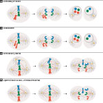
As chromosomes are partitioned during cell division, errors can occur along several pathways resulting in aneuploidy. |
Almost always. Cancer cells, which are overwhelmingly aneuploid, are an exception. They thrive and divide with a fury. The contradiction nagged Amon, an HHMI investigator, for years. How could aneuploidy—the kiss of death for most cells—be a hallmark in those cells with an unparalleled ability to proliferate? It didn’t make sense.
What was worse was that, outside of Down syndrome (a well-studied genetic disorder caused by an extra chromosome 21), aneuploidy was a mystery. Despite the prevalence of aneuploidy in cancer cells—more than 90 percent of solid tumors and about 75 percent of blood cancers display it—scientists had little understanding of how aneuploidy affects the physiology of a cell.
“It always bugged me,” says Amon. “So I decided to look into it.” Starting in 2002, Amon set out to determine the cellular impact of having an abnormal number of chromosomes and to resolve the aneuploidy paradox. Before long, she had a lot of company.
Today, a roster of experienced cancer researchers and molecular biologists is tackling aneuploidy’s effect on cells and its role in cancer. Their efforts are not purely academic: aneuploidy could be a useful target for new cancer drugs.

Leaning forward in a chair in her fifth-story office, Amon props her elbows atop a table, firmly clasps her hands, and declares, “I want to identify genetic or even chemical compounds that kill aneuploid cells preferentially.” Then, her voice growing soft, Amon describes watching her younger sister with breast cancer persevere through numerous rounds of treatment with Taxol, a common anti-cancer drug. “It’s brutal. If we could combine potential aneuploidy-selective compounds with Taxol, we could be more effective and lower the dose of Taxol. We could make the lives of people with cancer so much better.”
Extra Gain, Extra Pain
Amon’s first foray into aneuploidy was a shot in the dark. “I had a talented technician named Monica Boselli. She and I used to do crazy experiments all the time,” says Amon. In 2002, she asked Boselli to make a few aneuploid cell lines so they could get a good look at them—easier said than done. First, Boselli learned an arcane chromosome transfer strategy using molecular markers to select and move individual chromosomes from one yeast cell to another. She created 13 strains of haploid yeast, each with one or two extra chromosomes. Then, Boselli closely monitored the transplanted chromosomes, as yeast have a talent for ridding themselves of extraneous genetic material.
Boselli’s efforts worked, and she and Amon, along with Amon’s then postdoc Eduardo Torres, immediately observed that the extra chromosome was not dormant. In every case, the chromosome was active: hundreds of genes were being expressed from its DNA, and, Amon later confirmed, those gene transcripts were translated into proteins.

Next, Amon examined the appearance and activity of the aneuploid yeast. All 13 aneuploid cell lines, regardless of which of the yeast’s 16 chromosomes was added as the extra, shared common traits. They all had defective cell cycles: they multiplied more slowly—some couldn’t even form colonies—and many were 10-20 minutes slower to initiate cell cycle division than their normal, wild-type counterparts. This confirmed a common assumption that aneuploidy reduces a cell’s ability to reproduce normally. The yeast also exhibited increased glucose uptake and sensitivity to growth conditions that hindered protein folding and the breakdown of proteins.
All in all, the findings suggested that aneuploid cells require extra energy for survival, and that they struggle to fold and degrade proteins. The protein degradation pathways appeared to be working at full tilt trying to rid the cell of unfolded and misfolded proteins and parts of proteins. The subunits of many protein complexes are encoded across multiple chromosomes, so an extra or missing chromosome will result in isolated subunits with no partners. In fact, work published this April in Cell by HHMI Investigator Jonathan Weissman, at the University of California, San Francisco, showed that healthy cells produce equal amounts of the proteins that function together in a complex. Thus, any change in those carefully maintained ratios is likely to cause problems in a cell.
“It was pretty obvious [aneuploid cells] had major issues in the protein quality control department,” says Amon. She, Torres, and Boselli published the results in 2007. A few years later, Amon followed up with images of aneuploid yeast choked with clumps of fluorescently labeled protein aggregates shining like green warts in the cells.
Amon loves yeast: the smell of yeast, the pink and ivory colonies, the teeny bubble-like yeast spores. But after one too many colleagues insinuated that her aneuploidy findings were applicable only to yeast, Amon turned to mouse cells to determine if what they found in yeast was also true in mammals.
As she expected, the mouse cells had cell cycle defects and stressed protein control pathways. “It was the same,” says Amon of the mammalian work published in 2008 in Science. “Definitely the same.” A paper published this year by Zuzana Storchova and colleagues at the Max Planck Institute of Biochemistry in Germany confirms Amon’s findings, this time in human cells: aneuploid human cells, from a variety of sources, upregulate metabolic and protein degradation pathways.
Tug of War
Three miles south and across the Charles River, David Pellman, also an HHMI investigator and yeast aficionado, was simultaneously trying to figure out how aneuploidy affects the function of cells and obtain more details about how it occurs in the first place.
In addition to an abnormal number of chromosomes, most aneuploid cells have an abnormal number of centrosomes—small organelles that control the position and action of chromosomes on spindle fibers during cell division. Healthy cells have just two centrosomes during mitosis—division of somatic, or non-germ, cells—but aneuploid cells often have three or four. In 2008, at the Dana-Farber Cancer Institute and Boston Children’s Hospital, Pellman and colleagues began generating cell lines (a variety of mouse and human) differing only in centrosome number. At the time, it was well established that extra centrosomes generate aneuploidy, but the mechanism by which that occurred had not been directly observed.
Watching the cells by using a live imaging system, Pellman’s team found that cells with extra centrosomes divide into two, just like normal cells, yet the extra centrosomes produce additional spindle fibers that pull chromosomes in the wrong directions. This tug of war can result in lagging chromosomes that don’t make it to the correct side of the cell prior to division. The daughter cells, therefore, are aneuploid. “It was an unexpected mechanism,” says Pellman, but one that confirmed that centrosomes are active participants in chromosome segregation errors that lead to aneuploidy.
| An abnormal number of centrosomes—small organelles that control the position and action of chromosomes on spindle fibers during cell division—can cause aneuploidy. |
Aneuploidy can also be caused by a weakened or damaged spindle checkpoint, the security system in the cell that ensures chromosomes are accurately separated, says Hongtao Yu, an HHMI investigator at the University of Texas Southwestern Medical Center in Dallas. A host of spindle checkpoint proteins, such as Mad2 and BubR1, monitor the attachment of spindle fibers to kinetochores, sticky protein hubs at the center of chromosomes. In the case of incorrect fiber-kinetochore attachments, checkpoint proteins bind and inhibit a protein complex that would activate the next stage of cell division.
Complete loss of the spindle checkpoint wholly kills cells, says Yu. But lesser defects, such as low levels or decreased activity of Mad2 or BubR1, result in irregular attachments, continued cell division, and chromosome mis-segregation. “Subtle changes of the spindle checkpoint system allow cells to live, but cause aneuploidy,” says Yu.
| Defects in spindle checkpoint proteins can cause chromosomes to lag behind during cell division, as seen in these human cells. |
Paradoxically, it turns out that too much of a spindle checkpoint protein can also cause aneuploidy. Overexpression of Mad2 in mouse cells causes aneuploidy, a finding published by Rocio Sotillo, an HHMI international early career scientist at the European Molecular Biology Laboratory in Monterotondo, Italy, in 2007. The excess protein initially arrests cells in the phase of the cell cycle when the chromosomes are lined up along the cell’s equator. A few cells manage to “escape” that block, says Sotillo, and proceed with mitosis. But they overwhelmingly escape with baggage—extra or fewer chromosomes.
Other cell division defects and spindle checkpoint errors cause aneuploidy as well, and evidence from organisms such as Drosophila and plants reaffirms that the results of aneuploidy are slowed cell proliferation, sickness, and death. Unfortunately, these observations still failed to answer the question that nagged at Amon, Pellman, and others: if extra chromosomes reduce cell viability, how and why do aneuploid cancer cells thrive?
Elephant in the Room
In 1902, while observing the abnormal development of aneuploid cells in sea urchin embryos, German zoologist Theodor Boveri theorized that a tumor might develop from such a cell. Later scientists indeed found that most tumors are aneuploid, but many cast the finding aside as an oddity. For decades, the role of aneuploidy in cancer was largely ignored.
“If you look at old cancer reviews, they never put aneuploidy in there,” says Stephen Elledge, a geneticist and HHMI investigator at Harvard Medical School. “It has always been the elephant in the room.” That may be because, during the 1970s and 1980s, it was significantly harder to identify the cellular effects of a whole additional chromosome, as opposed to a single oncogene, Elledge suggests. “It just hurt people’s heads.”
With the advent of new imaging and sequencing tools beginning in the early 1990s, aneuploidy received renewed attention—but soon became stymied by controversy. Leading cancer researchers suggested aneuploidy was simply a by-product of cancer, an unimportant consequence. Others vehemently argued that it could be the sole cause of cancer. “There was a huge debate,” says Pellman. But it didn’t scare him away from the field.

Prior to working full time in the lab, Pellman was a pediatric oncologist. Like Amon, a view through a microscope drew him to the study of aneuploidy: over and over, he observed abnormal structures in the nucleus that indicated aneuploidy in the cells of his young cancer patients. “I spent a lot of time looking at these abnormal nuclear structures,” says Pellman. “There was an obvious connection.”
So he decided to put one aspect of Boveri’s hypothesis—that whole genome duplication, which causes high rates of aneuploidy, might cause cancer—to the test. Pellman instructed one of his postdocs, Takeshi Fujiwara, to produce tetraploid (twice the normal chromosome number) mouse cells, which have a high rate of chromosome mis-segregation and thus rapidly become aneuploid. Then, Fujiwara was to inject those cells into mice. Although Pellman thought it was important to test the idea, he felt it was almost too simple, “too good to be true,” for the tetraploid cells to cause cancer. Fujiwara’s initial results showed that the cells indeed caused aggressive tumors in the mice. “It was what we were after, but it was nevertheless very surprising that it worked so well,” Pellman recalls. The discovery provided strong evidence that tetraploidy, and the resulting aneuploidy, promotes tumor development.

Since then, Pellman’s lab has been working to determine how aneuploidy might cause tumors. In 2012, his team showed that inside micronuclei—small membrane-bound bodies that partition extra chromosomes away from the cytoplasm—chromosomes are subject to extensive DNA damage. This suggests that one way aneuploidy causes cancer is the old-fashioned way: through mutations that activate cancer-causing genes or inactivate tumor suppressors. Additionally, Pellman’s lab recently discovered evidence showing that extra centrosomes, independent of generating aneuploidy, can lead to the activation of Rac1, a protein involved in cancer cell signaling and tumor invasion (published June 5, 2014, in Nature).
More evidence is mounting that aneuploidy promotes cancer by altering oncogenes and tumor suppressors. Last year in Cell, Elledge and colleagues at Harvard reported that cancer genomes select for extra chromosomes that contain potent oncogenes, and select against extra chromosomes rich in tumor suppressors (see “The Method in Cancer’s Madness,” Spring 2014 HHMI Bulletin). At the Mayo Clinic in Minnesota, molecular biologist Jan van Deursen and colleagues have found that aneuploidy predisposes mice with only one working copy of p53, instead of a normal pair, to lose that copy of the suppressor gene and develop tumors.
Based on current understanding, aneuploidy appears to promote tumor production through multiple mechanisms, but not by increasing a cell’s ability to multiply. That, at least, helps resolve the aneuploidy paradox. “We think [aneuploidy] offers tumor cells something other than proliferation, something else that is good for tumorigenesis,” says Amon. (See sidebar, “Stressed-Out Cancer Cells: The Benefits of Aneuploidy.”)
|
WEB EXTRA Learn why an abnormal chromosome number might give cancer cells an edge. |
But does aneuploidy cause cancer on its own? Van Deursen’s work suggests that it does not. For more than a decade, van Deursen has been engineering mice to produce low levels of individual spindle checkpoint proteins, including Bub1 and BubR1 (complete loss of such proteins is lethal to embryos). Some of the mice develop cancer, from lung cancer to lymphoma to liver tumors, but not all. What’s more, van Deursen can’t predict which checkpoint protein that he knocks down—and he has tried a dozen—will result in a tumor.
“If it were merely aneuploidy that drives malignant transformation, then we’d expect that all the mice would have cancers, and many cancers. But that really isn’t the case,” says van Deursen. “It’s likely the answer is aneuploidy plus something else. So you will have two, three, or four independent, tumor-promoting activities.”
Today, most researchers, including Amon, agree that oncogenes are the main drivers of cancer, and aneuploidy contributes variation and instability to the tumor cells. If oncogenes are the spark, aneuploidy is the kindling. “We’re at the point where there’s a pretty strong trust that aneuploidy is driving cancer,” says Elledge. “The question is, How potent is it?”
Targeting Stress
Until that question is answered, most aneuploidy research remains focused on basic mechanisms. But a few inquisitive scientists are dipping their toes into translational research, looking for ways to target or exploit aneuploidy to preferentially kill cancer cells.
“Aneuploidy is definitely stressing the cells, and the cells must depend on stress support pathways more than normal cells do,” says Elledge. “So if you can tinker with that, you can potentially make the cell vulnerable or kill it. This could certainly augment other therapies.”

Some of Pellman’s work suggests that targeting cells with too many centrosomes might be a way to selectively target cancer cells. Several drug companies are pursuing small molecule drugs to inhibit a specific motor protein that Pellman identified as essential for the survival of extra centrosome-containing cancer cells.
At UT Southwestern, Yu and colleagues recently completed an experiment in which they silenced each of the 20,000+ genes in human cancer cells, one by one, and then treated the resulting cells with Taxol. Their goal was to see which genes help cells avoid death by Taxol, which is believed to cause massive aneuploidy, and which genes promote Taxol-triggered death. Many of the genes they identified encode components of the spindle checkpoint, suggesting potential drug targets to help Taxol work better, says Yu. Those targets could lead to the kind of drugs Amon has long dreamed of, to improve the effectiveness of current chemotherapy regimens and reduce side effects.
Back at MIT, Amon’s translational work is in full swing. In collaboration with a Harvard screening facility, Amon has been searching through libraries of compounds for chemicals that preferentially prevent aneuploid cells from multiplying. She has identified several. One of them is a chemical involved in the formation of lipids that appears to target highly aneuploid cells.
Amon’s team found that by combining Taxol with the new compound at concentrations where each could kill 10 percent of cancer cells if given separately, the mixture was able to destroy cancer cells with an efficacy of 90 percent. “Now we’re working to treat mouse models of cancer and determine the mechanism,” she says. “We’re really excited about it.”
