Stay Connected. Manage Your Care.
Access your health information anytime and anywhere, at home or on the go, with MyHealth.
- Message your clinic
- View your lab results
- Schedule your next appointment
- Pay your bill
The MyHealth mobile app from Stanford Health Care puts all your health information at your fingertips and makes managing your health care simple and quick.
Guest Services
24/7
We are available to assist you
whenever you need it. Give us a call at
650-498-3333 or
PHYSICIAN HELPLINE
Have a question? We're here to help! Call 1-866-742-4811
Monday - Friday, 8 a.m. - 5 p.m.
REFER A PATIENT
Fax 650-320-9443
Track your patients' progress and communicate with Stanford providers conveniently and securely.
Stroke Diagnosis and Evaluation
A stroke occurs when blood vessels carrying oxygen to the brain burst or become blocked, damaging the brain cells' ability to control sensation, movement, or function, and eventually causing the nerve cells to die.
The ability to pinpoint quickly the precise location of a stroke and determine the extent of damage is critical in making treatment decisions during a stroke emergency. For instance, the doctor must be able to quickly determine whether the stroke is ischemic (arising from a blocked blood vessel) or hemorrhagic (bleeding caused by bursting of a blood vessel) before the appropriate therapy can begin.
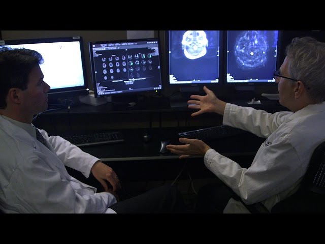
State-of-the-art brain diagnostic devices
The Stanford Stroke Center is one of the few places in the country that has more than a dozen state-of-the-art brain diagnostic devices available to obtain in-depth information about a patient's status.
These highly sensitive tools are of critical importance in diagnosing abnormalities that place you at high risk for stroke, such as a blocked blood vessel or the presence of an aneurysm or AVM.
Diagnostic tests
To obtain complete diagnostic information, several of the following diagnostic tests may be performed during an evaluation for stroke or stroke risk.
-
Computerized
tomography (CT) scan
This is generally the first diagnostic test done after a patient with a suspected stroke arrives in the emergency room. It is used to quickly distinguish between ischemic and hemorrhagic strokes. The test involves the use of low-dose X-rays to visualize the brain.
-
Magnetic
resonance imaging (MRI)
MRI is an advanced diagnostic tool that provides a high level of anatomic detail for precisely locating the stroke and determining the extent of damage. Due to its high level of sensitivity, MRI is especially useful when the stroke involves small blood vessels.
The technology involves use of a strong magnetic field, and is performed in a special room free of metallic equipment. Recently, there have been great advances in the early detection of stroke using diffusion (DWI) and perfusion (PWI) weighted imaging. These methods allow early and more accurate detection of acute stroke, improving our ability to treat patients with cerebrovascular problems. Stanford has been a leader in the development of this technique.
-
Magnetic
resonance angiography (MRA)
MRA is a noninvasive technology for imaging the cerebral blood vessels, and yields valuable information regarding blood supply to the brain. The use of intravenous contrast agents has provided great improvements in accurately viewing the cerebral blood vessels. Many such techniques have been pioneered by researchers at Stanford.
-
Transcranial
doppler (TCD)
TCD a new, noninvasive ultrasound procedure that allows the assessment of blood flow through the cerebral vessels via a small probe placed against the skull. TCD is a portable test that can be performed frequently at the patient's bedside to follow the progress of medical treatment for stroke.
-
Carotid
duplex scanning
This is a noninvasive study to diagnose blockage in the carotid arteries. This technology involves recording sound waves that reflect the velocity of blood flow.
-
Radionuclide
SPECT scanning
This provides data on relative blood flow using the radionuclide Technetium99.
-
Positron
emission tomography (PET) scanning
A PET Scan measures brain cell metabolism, can determine if brain tissue is functioning even if blood flow to that area appears to be diminished.
-
Cerebral
angiography (angiogram)
This method requires injection of a contrast dye through a major artery (usually the femoral artery in the thigh) for evaluation of blood flow to the brain. This procedure is completed in Stanford's Cath/Angio lab. The procedure time is approximately two to three hours; bed rest for six hours is required after the procedure.
-
Transesophageal echocardiography
This involves placing a flexible tube in the esophagus (tube to stomach) to directly image the heart and assess its function and structures.
-
Ultrasound
An ultrasound is a diagnostic test that uses high frequency waves of sound to help examine the movement, size and shape of blood vessels.
Clinical Trials
Clinical trials are research studies that evaluate a new medical approach, device, drug, or other treatment. As a Stanford Health Care patient, you may have access to the latest, advanced clinical trials.
Open trials refer to studies currently accepting participants. Closed trials are not currently enrolling, but may open in the future.
Our Clinic
The Stanford Stroke Center is a pioneer in using the latest surgical techniques and innovative therapies to rapidly treat individuals experiencing a stroke.
