Skip to content Skip to navigation

Home

Technologies Offered:
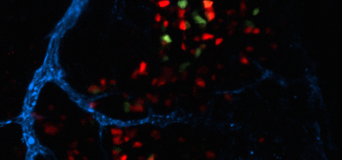
Light Microscopy

  • Confocal microscopy (scanning and spinning-disk)
  • Two-photon microscopy
  • Wide-field fluorescence microscopy
  • Digital deconvolution
  • Transmitted-light imaging (phase, DIC, histology)
  • High-content screening (Confocal and Wide-field)
  • Super-resolution imaging (STORM, STED and SIM)
  • Cell surface imaging with <100 nM z-resolution (TIRF)
  • Specialized microscopy (FRAP, FRET, FCS, FLIP...)
  • Fluorescence lifetime imaging microscopy (FLIM)
  • Image Analysis expertise
  • Software for image analysis, processing, and deconvolution
    • Imaris, Volocity, SVI Huygens, SoftWoRx. Microvolution

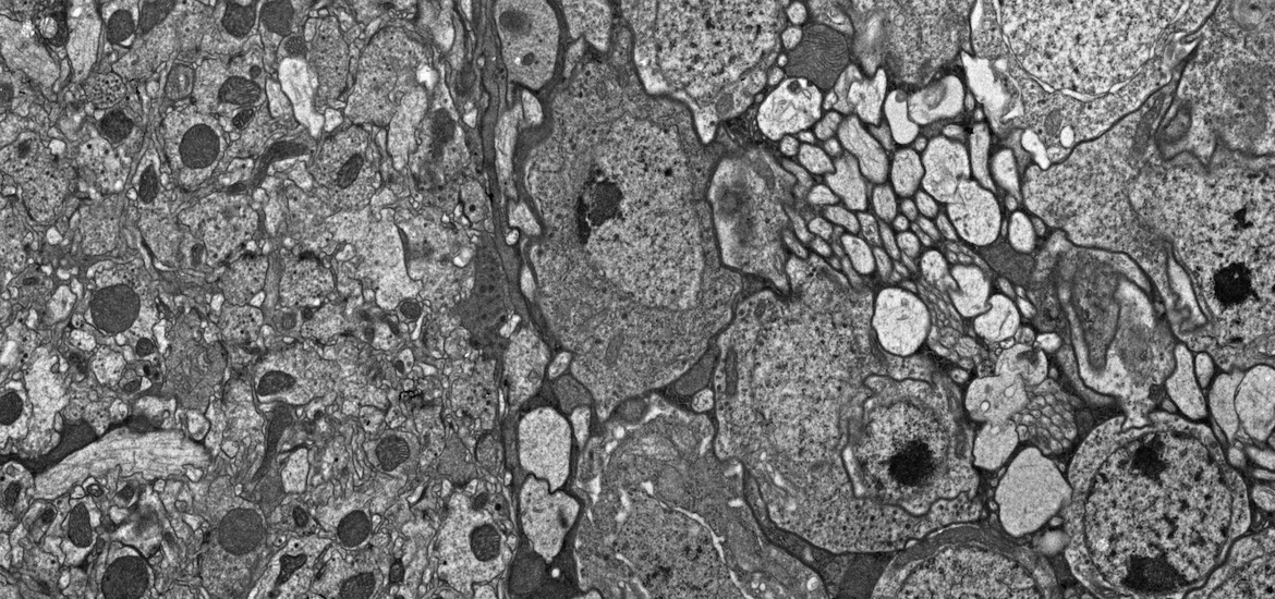
Electron Microscopy

Transmission Electron Microscopy (biological)

  • Chemical and cryo-fixation processing
  • immuno-EM
  • Correlative LM to EM: CELM
  • negative staining
  • ultramicrotomy, cryo and plastic
  • electron tomography (plastic sections)

Scanning Electron Microscopy

  • variable pressure/environmental
  • high resolution FE-SEM
  • Immuno-SEM
  • array tomography SEM, CLEM

The Cell Sciences Imaging Facility (CSIF) is a Beckman Center supported, university service center that provides high resolution, state-of-the-art light and electron microscopy technologies for imaging and analyzing the molecular and structural organization of cells, tissue and bioengineered materials.  The CSIF operates two sites at Stanford University: The SOM Beckman Center CSIF and the SOE Shriram Center CSIF.  Both facility sites are open to all members of the Stanford community as well as to external academic and industry researchers (with approval of the facility Director).


Events

CSIF SEMINAR: Why Cryo EM for Cells?
Friday, February 26, 2016 - 12:00

CSIF SEMINAR: Why Cryo EM for Cells?

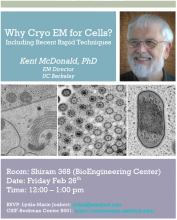
Including Rapid Techniques for Cryo Processing of Biological Specimens.


Speaker: Kent McDonald PhD, EM Director UC Berkeley.

Room: Shiram 368

Date: Friday Feb 26th

Time: 12:00 – 1:00 pm RSVP: lydiaj@stanford.edu (Lydia-Marie Joubert)


Reading Material: 1. McDonald, K. 2014. Protoplasma. 251(2): 429-48. http://www.ncbi.nlm.nih.gov/pubmed/24258967

2. McDonald, K. 2014. Microsc Microanal. 20: 152-163. http://www.ncbi.nlm.nih.gov/pubmed/24252586


TABLE TOP SEMs

Hitachi TM3030Plus-Oxford EDS

Hitachi Atmoscpheric SEM

Both SEM models will be on display in CSIF-­‐Shiram (Room 023A): 22 ­‐ 26Feb

Please, sign up on the following doodle for a demo using your own samples:

http://doodle.com/poll/iuvcrf546bdemqkpuiuyqfgn/admin#table


MicAO - Adaptive Optics
Monday, February 22, 2016 (All day)
Starting Monday the 22nd of February, we will be hosting "MicAO", for 1 week, an adaptive optics solution for our spinning disk confocal in room B023 in the Shriram Center.
 
The system allow a significant increase in your signal (up to 100% at 20um) by correcting aberrations.
As it will be a significant improvement of our actual Spinning Disk I recommend anyone who is interested to demo the system, ideally with your own sample, and to give us feedback.
 
If you are interested please sign up on the following doodle:
 
 
If you want to learn more about the technology, Vincent from Imaging Optics will present MicAO, Monday the 22nd of February from 2:30pm to 3:3pm in room 054 in the Shriram Center.