 |
|
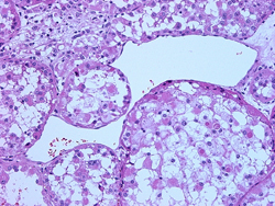
| Sinusoidal Vascular Pattern in Alveolar Soft Parts Sarcoma | |
Consulting Services
We provide the full range of diagnostic services for wet tissue specimens and glass slides sent in consultation. Our services include:
Glass Slides and Wet Tissue
- Stanford Surgical Pathology Faculty have served as consultants to pathologists, other physicians and patients for nearly 40 years. Cases submitted in consultation are reviewed by fellows and one or more members of the General Surgical Pathology faculty or subspecialty faculty such as cytopathology, dermatopathology, hematopathology and neuropathology.
- Fixative filled specimen containers are supplied to the inpatient wards, the outpatient clinics and the surgical suites of Stanford University Hospital and Clinics and Lucille Packard Children’s Hospital and to numerous outreach clients. Daily courier service is available within much of the Bay Area. We strongly encourage the submission of appropriate clinical history and laboratory data.
- Preliminary interpretation is typically available 24-36 hours from receipt of the specimen. Large specimens may require overnight fixation.
- In our interpretation of the specimen, we may resort to the judicious use of ancillary techniques. These may include immunohistochemical stains, flow cytometry, molecular pathology, cytogenetics, immunofluorescence, in situ hybridization, and electron microscopy.
- Critical and/or unexpected diagnoses will be immediately called to the referring clinician.
Immunohistochemistry
- Our immunohistology laboratory is fully equipped to perform a wide array of antibody stains. We practice judicious use of special techniques and do not apply them indiscriminately.
Immunofluorescence
- We perform direct immunofluorescence on tissue samples for antibody mediated rejection and subtyping of amyloidosis.
- Please submit tissue for direct immunofluorescence in Zeus or Michels transport medium. Zeus transport medium is supplied by our laboratory, (link) Once in transport medium, tissue is stable for several weeks at room temperature and can be picked up or shipped with your routine formalin fixed specimens.
Electron Microscopy
- Specimens for electron microscopic examination are preserved best in fixatives such as glutaraldehyde (link to supply). The tissues should be cut into 1 mm cubes and immediately placed in the fixative.
- Electron microscopy is rarely utilized in the diagnosis of neoplasms. Electron microscopy can be useful in the diagnosis of various metabolic disorders, amyloidosis, microvillous inclusion disease, Whipple’s disease, and medical renal diseases.
- Electron micrographs can be viewed in a variety of formats including hard copy and electronic.
Affiliated Services
- The Molecular Genetics Diagnosis Laboratory is directed by Drs. Iris Schrijver and James Zehnder. The laboratory performs gene rearrangement studies for T- and B-cell clonality by PCR, tests for microsattelite instability and PCR or FISH for chromosomal translocations.
- The Cytogenetics Laboratory is directed by Dr. Athena Cherry.The laboratory performs fluorescence in situ hybridization (FISH) studies for chromosomal translocations and rearrangements that are associated with particular types of leukemias/lymphomas and solid tumors.