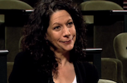


In March 2013, Lillian Fritz-Laylin packed a cooler full of human immune cells and hopped on a plane from San Francisco to Virginia. Once she arrived, she spent almost every day in a small, dark room in Eric Betzig’s lab at HHMI’s Janelia Farm Research Campus, watching the cells crawl across a microscope’s stage. A month later, she returned to the West Coast with more than 10 terabytes of data and information that would change the way a whole field looks at cell movement.
Fritz-Laylin, a postdoctoral researcher in HHMI Investigator Dyche Mullins’ lab at the University of California, San Francisco, studies “fast” immune cells, which zip around about 100 times quicker than most other cells. They need this speed to respond to infections and other problems before things get out of hand. She’d like to know how they get around so swiftly. Unfortunately, microscopes that can capture the details of cells moving at this speed are few and far between. Except in Betzig’s lab. He’s built several microscopes that can visualize live cells, up close, in three dimensions. An avid collaborator, Betzig invited Fritz-Laylin to bring her cells to his lab.
What the microscope revealed was striking. A typical cell moves by oozing: extending a thin surface-attached edge, while simultaneously retracting its backside. The fast cells, as seen moving in 3-D, were different. They were covered in large, dynamic projections that extended and retracted in all directions. Fritz-Laylin suspects the fast cells are using forces produced by the rear retractions—like a tube of toothpaste being squeezed from the bottom.
| Fast immune cells move by extending large, dynamic projections in all directions. Video courtesy of the Mullins Lab. |
The Janelia Visitor Program welcomes scientists such as Fritz-Laylin from around the world to the Ashburn, VA, campus to do research. To date, more than 180 visiting scientists from the United States and 23 other countries have participated in the program since it launched when Janelia opened in 2006.
Some collaborations last a few weeks; others go on for years. Some involve huge endeavors like mapping all the nerve cells in the fly brain. Others tackle smaller problems like sleuthing out the neurons that affect hunger in mice. By bringing together people with different expertise, the collaborations accomplish more than a single lab could have done on its own.
“Scientists come here and get access to equipment, budget, scientific reagents, and the ability to do things they couldn’t do at their home institutions,” says Janelia Executive Director Gerry Rubin. “It’s a very special opportunity that doesn’t really exist elsewhere.”
“We really want to make sure that our state-of-the-art instrumentation or any new technique that we’ve developed can reach the community at large,” explains Science Program Manager Zarixia Zavala-Ruiz.

Fly Fighters
One of the very first visitors to Janelia was longtime HHMI Investigator David Anderson. His California Institute of Technology lab group is trying to understand how the brain processes emotional behavior such as fear, anxiety, and aggression. Over three years, Anderson traveled to Rubin’s lab every couple of months to develop an assay to learn whether the fruit fly Drosophila melanogaster experiences fear.
“I would spend a week here and there, buried in the laboratory, running my experiments,” Anderson recalls. “I really enjoyed the opportunity to get away from my administrative responsibilities at Caltech and immerse myself in doing bench science with my own two hands.”
Anderson’s initial project grew into a second, larger one focused on aggression. His postdoc, Eric Hoopfer, moved to Janelia to carry out the five-year project, which centered on a collection of 8,000 transgenic Drosophila lines. Created by Rubin and his team, each line has a different set of nerve cells that can be turned on and off. The entire collection covers the estimated 150,000 neurons that make up the central nervous system in the adult fruit fly. At the time, the lines were an extremely hot commodity for scientists interested in nerve cell function.
| Eric Hoopfer is systematically turning on sets of neurons in the fly brain to transform normally docile flies into aggressive fighters. Video courtesy of the Anderson Lab. |
Hoopfer systematically turned on sets of neurons in the fly brain hoping to transform normally docile flies into aggressive fighters. “In the vast majority of days, they’d do nothing,” he admits. But once in a while, the flies would start going after each other.
“There are very specific populations of neurons that are involved in aggression, and looking for them is like looking for a needle in a haystack,” Hoopfer explains. He ended up screening about 3,000 fly lines. Twenty of those lines contained neurons that increased aggression.
Back at Caltech now, Hoopfer is writing up his results and starting to figure out how the labeled neurons contribute to aggressive behavior. “The ability to do this project has been a great help to my career,” he says. “The lines that I found are basically tools that I’ll be able to use to start my own independent research group. There are years of work to be done figuring out how just these 20 lines work.”
| Fight Club for Flies: Science Take with James Gorman |
Hunger Circuitry
Janelia Group Leader Scott Sternson also likes to collaborate. Even before he set up his lab at Janelia, he had a list of scientists he wanted to work with. So it was perfectly natural for him to propose a project with neuroscientist Anirvan Ghosh within a few hours of meeting him.
Sternson studies hunger. Back in 2010 when he first met Ghosh, he was walking neuron by neuron through the paths that sense when a mouse’s brain needs fuel, hoping to create a map of the circuitry that controls hunger. Unfortunately, the circuits weren’t linear. Instead, the nerve cells’ axons—the finger-like regions that transmit signals to neighboring neurons—branch into different parts of the brain, making it hard to figure out which fork in the road to follow. Sternson knew he could determine the correct path by blocking the axons’ output points, called synapses, and seeing if that made the mouse hungry. But he needed help targeting the synapses.
| Thomas Jessell of Columbia University explains what happens at a synapse. |
That’s where Ghosh came in. He was studying the molecular biology of synapses at the University of California, San Diego. Sternson realized that by collaborating, they might be able to build a “circuit breaking tool” that could home in on selected synapses and turn them off. The pair decided to base their tool on a molecule called hM4D, which turns off nerve cells in the presence of a drug called clozapine-N-oxide (CNO).
For the project, Ghosh recruited Tev Stachniak, a postdoctoral fellow from McGill University in Montreal who was interested in neural circuits. With guidance from both Ghosh and Sternson, Stachniak tinkered with hM4D and eventually got it to specifically target the synapses of interest. The effects of CNO last about an hour, just enough time to test the circuit breaker’s impact on mouse feeding behavior.
The project, which is winding down, was a great success. Stachniak discovered that part of the hunger pathway involves neurons that mediate reward-motivated behavior in general, hinting at the existence of a core circuit shared by these behaviors. The tool, described in a publication in press at Neuron, can be used to target synapses involved in other circuits as well, making it widely useful for neuroscientists. Ghosh, who moved to Roche as the Global Head of Neuroscience Discovery in 2011, is starting to think about using similar tools in more complex animals such as primates. And Sternson, of course, is already on to his next collaboration.
Brain Maps
The only way Group Leader Albert Cardona will accomplish his ambitious project is if he engages lots of visiting scientists. He wants to create a neural wiring map of the fruit fly larva brain. That’s about 12,000 neurons, 3,000,000 synapses, and 50 person-years of work. So far, he’s brought in 39 scientists from 19 labs around the world. He teaches them how to trace the neurons in electron micrographs created by Rick Fetter, a principal scientist at Janelia, from a Drosophila larva that was sliced into about 5,000 sections, each 50 nanometers thick.

One of his visitors is Katharina Eichler, a PhD student from Andreas Thum’s lab at the University of Konstanz in Germany. She’s been working with Cardona for about six months, spending her days hunched over a computer and clicking a cursor on the black-and-white micrographs. She expects to be doing this for at least another six months.
Eichler and the other visiting scientists do their mapping using a Web application called CATMAID (Collaborative Annotation Toolkit for Massive Amounts of Image Data) that is like a Google Maps for the brain. The roads—neurons, in this case—are already on the map. “It’s just like the OpenStreetMap.org project that asks contributors worldwide to annotate the place they live in with their knowledge of the local geography,” says Cardona. Once the scientists learn the program, they can log in from their own computers and trace neurons from anywhere in the world.
Most of the scientists stay with Cardona for one to six months, learning how to map the fly brain. Eichler is an exception because she’s decided to complete her PhD at Janelia in collaboration with the University of Konstanz. Eichler could have done the project in Germany, but, she explains, “I think it’s important to be here because it’s really helpful to be able to talk to people who have worked with a certain neuron or area of the brain before.”
Eichler’s studying the mushroom bodies—a pair of structures involved in olfactory learning and memory. By the time she leaves Janelia, she hopes to have traced all of the approximately 320 nerve cells that make up the structures.

So far, Eichler and the rest of the team have mapped about 12 percent of the nervous system. But this is not the best measure of their progress. “Biologically speaking, there are so many questions you can answer with a fraction of a reconstruction,” says Cardona. “You don’t have to finish the whole thing to extract enormous value. You extract value as you go.” Eichler has already proven this: even though she’s only about halfway through her mapping, she’s already showing how the connections between the mushroom body neurons contribute to the various roles the structure plays in the fly brain.
Massive Sequencing Experiments
Sacha Nelson and his postdoc Ken Sugino had a big idea that needed some big resources. Resources like immense computational power and instrumentation that could only be found at a place like Janelia.
The pair had figured out how to isolate genetically similar cells from a mouse’s brain, pool the RNA from the cells, and determine the RNA’s sequence. The result was a transcriptional profile—a unique fingerprint—that could be used to distinguish different cell types. If they could create a database of RNA produced by each of the cell types in the mouse brain, it would be an incredible resource for neuroscientists.
“Ultimately, if this database were big enough, any scientist could just look up a favorite neuron and read out that neuron’s profile,” explains Janelia Group Leader Adam Hantman. That information could help the scientist figure out what the neuron does and how it does it.
The problem was that, by Sugino and Nelson’s estimate, the brain has between 5,000 and 10,000 different cell types. Sequencing each of those cell types would require more resources than were available in a small academic lab such as Nelson’s at Brandeis University. So Nelson started casting around for collaborators. “I looked for people at Janelia,” he says, “who were interested in using the scale available there to do the massive sequencing experiments that were not practical to continue in my own lab.”
That’s how they teamed up with Hantman and Janelia Group Leader Sean Eddy. Hantman needed help characterizing some mouse brain cells he was studying. Eddy, on the other hand, was interested in the genomic implications of the project. “It would be really cool if you could get each cell type and say, ‘This is the genetic program this guy is running, and this is the program that guy is running,’” he explains. “Then you could start to ask how transcription regulation works, how it evolved, and how is it different between species.”
Eddy also happened to have two postdocs, Lee Henry and Fred Davis, who were working on a similar technique to purify fly brain cells and sequence their RNA. Thus, the big project got even bigger. The new goal was to sequence all the cell types in both the mouse and fly brains.

As word got out about the project, many Janelia scientists approached the team, asking them to create RNA profiles for the mouse and fly brain cells they were studying. As a result, in September 2012—one and a half years after it started—the visitor project morphed into a larger-scale, permanent project called NeuroSeq.
Sugino, Henry, and Davis are now full-time employees at Janelia on the NeuroSeq team. Nelson is back in his lab at Brandeis, overseeing the project from afar. The team is also spending one-third of its time helping Janelia scientists ask specific questions about brain cells. Sternson has already approached them to learn how changes in feeding affect the RNA produced by certain cells in the mouse brain.
A New Adventure Weekly
Janelia Group Leader Eric Betzig calls the Visitor Program his “secret weapon.” Without it, he wouldn’t be able to share the cutting-edge, but very large, immobile microscopes he builds. People have to come to him. “We need to show that the tools are useful,” he explains. “And I need outside collaborators to do that.”
One of his creations is the Bessel beam microscope that Fritz-Laylin used. It’s a hulking assembly of lasers and lenses that uses a thin sheet of light—similar to a scanner at a checkout counter—to acquire tens of thousands of images from a living specimen. By piecing together the frames, Betzig can assemble dazzling three-dimensional movies that show the inner workings of cells.
In 2012 and 2013, Betzig averaged about 20 visitors per year. A quick glance at his calendar for January 2014 shows four collaborators visiting back to back: a researcher from Duke University, a scientist from the National Institutes of Health, a collaborator from Johns Hopkins University, and finally, a group from Harvard University.
Typically, visitors—who range from individual scientists to teams of specialists—arrive on Sunday and jump into data collection first thing Monday morning. The microscopes are complicated enough that they have to be operated by Betzig’s postdocs, who are willing to work 18-hour days to make sure the collaborators leave with as much data as possible.
Betzig will say yes to any collaboration that could benefit from his microscopes. “The variety of things we’ve looked at is all over the map,” he says. “We’ve gone from bacteria up to huge fish, and everything in between. Every week is a new adventure.” Two visitors—one from Harvard and one from the University of California, San Francisco—are even using the Visitor Program to build their own light sheet microscopes in collaboration with Betzig and Janelia’s Instrument Design & Fabrication team.
Adding On
“I think everybody benefits from the Visitor Program,” says Janelia Scientific Program Director Ulrike Heberlein. “Janelia benefits from having really wonderful and smart people contribute to the intellectual discourse. The visitors benefit from being in this really fantastic environment where they can try things they otherwise might never be able to do.”
When Heberlein first came to Janelia two years ago, she thought the Visitor Program might benefit from a little more structure. “I wanted to have more rules and criteria for what gets funded and what doesn’t,” she explains. She soon shelved that idea. “It turns out that the real beauty of this program is the flexibility,” she says.
Rather than changing the program, Heberlein has decided to add to it. This spring, Janelia launched its Visitor Graduate Fellowship. Aimed at students from around the world, the program will allow the young scientists to create collaborative projects that will become part of their thesis research.
And, like the Visitor Program, the sky is the limit. Even longshot projects find a home at Janelia.
“We’re willing to say, ‘Well, this [visitor project] may only have a 20 percent chance of working, but if it works it’s going to be really transformational, so we’ll do it,’” says Rubin. “We have a high-risk, high-reward attitude toward these projects.”


