


As a fish swims, nerve cells fire in its brain, sending signals racing along a neural network ending in muscles that make its fins flap and its tail swish. By using a molecule called CaMPARI to permanently mark neurons as they fire, scientists can now watch as signals light up such neural networks in live animals.
CaMPARI came out of a collaborative project spearheaded by Eric Schreiter, a senior scientist in Group Leader Loren Looger’s lab at the Janelia Research Campus. The team started with a protein called Eos, which emits a green glow until it’s exposed to violet light. The light changes the molecule’s structure, causing it to glow red. By combining Eos with the calcium-sensitive protein calmodulin, the researchers were able to couple Eos’s color change to the burst of calcium that accompanies neuronal signaling. The resulting molecule indelibly tags firing neurons with a red glow in the presence of violet light.
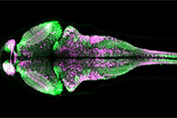
| A snapshot of zebrafish brain activity captured using CaMPARI. The microscope scans through a four-day-old zebrafish brain show neurons that were more active (magenta) or less active (green) while the fish was freely swimming. |
“Ideally, we would flip the [violet] light switch on while an animal is doing a behavior that we care about, then flip the switch off as soon as the animal stops the behavior,” Schreiter explains. “So we’re capturing a snapshot of neural activity that occurs only while the animal is doing that behavior.”
The scientists published their results February 13, 2015, in Science. Although they are still tinkering with CaMPARI to make it more sensitive and reliable, they’ve already made it available to scientists on Addgene and the Bloomington Drosophila Stock Center. Janelia Group Leader Misha Ahrens is also distributing CaMPARI-expressing zebrafish.