


Eric Betzig is a physicist and an engineer: he thinks in terms of light waves and energy, and when he tinkers in the lab, it is with lasers and mirrors and beam splitters. He’s the first to admit that he is no biologist. But lately, his lab at the Janelia Research Campus has been bustling with experimental biologists who arrive toting vials of cells and leave with gloriously detailed videos of molecular life in motion. Those scientists return to labs all around the world with their minds racing, full of ideas about how Betzig’s newest invention—a microscope that lets users collect high-resolution, three-dimensional images over prolonged periods—might transform their research.
It’s not the first time biologists have clamored to get their hands on one of Betzig’s groundbreaking imaging tools. New technologies have streamed steadily out of Betzig’s lab as he has resolutely ticked off items on the ambitious to-do list that he laid out when he came to Janelia in 2005: faster microscopes, gentler microscopes, microscopes that look deeper, and microscopes that reveal more detail. Nine years later, he says, “I’m getting close to done.”
Biologists are accustomed to designing their experiments around the limitations of available tools. For fluorescence microscopy—the method of choice for finding and following specific molecules inside cells—that has in the past meant choosing between imaging speed, detail, or the well-being of the sample. But Betzig has always been eager to hear about his colleagues’ frustrations. “If you know the problem, that’s half the battle to coming up with the solution,” he says. “That’s the source of creativity.” Betzig has spent the past decade applying his creativity to finding ways around the usual imaging trade-offs, thus giving biologists the freedom to apply their creativity to investigating the questions they hope to answer.

In October, Betzig’s creativity was recognized with the 2014 Nobel Prize in Chemistry; he shares the prize with Stefan Hell of the Max Planck Institute for Biophysical Chemistry in Germany and William Moerner of Stanford University. The prize committee lauded the three scientists for their development of super-resolved fluorescence microscopy—methods of visualizing objects so small that, until recently, distinguishing them with a light microscope was considered a feat that would defy the fundamental laws of physics.
Living Color
Betzig helped launch a revolution in super-resolution microscopy in 2006, when he developed a method that he and his collaborator Harald Hess called photoactivated localization microscopy, or PALM. The technique creates stunningly detailed images of cells by taking advantage of fluorescent labeling molecules that can be switched on and off with a pulse of light. In PALM, a sample labeled with these fluorescent tags is imaged many times, with a small subset of the fluorescent tags switched on each time. Because just a smattering of molecules is glowing in the image, each one can be pinpointed with precision. Compiling thousands of images yields a picture in which nearly all fluorescently labeled molecules show up as individual and distinct.
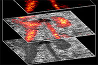
| Eric Betzig and Herald Hess explain how they joined forces to develop the first super-high-resolution PALM microscope. |
The Janelia campus—where both Betzig and Hess are now group leaders—was still under construction when Betzig first learned of the fluorescent probes that would make PALM possible. So he and Hess built a prototype of the microscope in Hess’ living room in La Jolla, California. Later, they set up shop in a trailer on the campus of the National Institutes of Health, where they worked with cell biologist Jennifer Lippincott-Schwartz and others to generate the microscope’s first spectacularly revealing images of cells at work. “The first time we put a dilute suspension of these [fluorescent] molecules on the surface, and we turned on the photoactivating light, we knew we had it,” he remembers. “It felt like flipping a switch.”
Betzig and postdoctoral researcher Hari Shroff “lived and breathed PALM” during his first few years at Janelia, adding features that made the technique more powerful, such as the ability to detect multiple colors—meaning more than one kind of protein can be tracked at once—and the possibility of imaging living cells. But Betzig knew the method had its limits. “It’s too slow, and you throw too much light at the cells,” he says, explaining that generating a single PALM image subjects samples to toxic light exposure time and time again. Because of that, Betzig realized, the principles of PALM would never translate into what biologists really craved: the same extraordinary resolution in three-dimensional images of living cells.
With a spate of other labs by then working to enhance super-resolution microscopy, Betzig decided it was time to move on. “Everybody and his kid sister was doing super-resolution then. You have to go to the areas where other people aren’t,” he says. “Those are the most fertile areas to find something useful.”
Fertile Fields
He saw plenty of opportunities. Now that he was at Janelia, mingling daily with scientists working at the forefront of neuroscience research, he became intent on improving their capability to look inside the brain. To investigate how nerve cells mediate thoughts and actions, scientists need visual access to all parts of the brain—but most microscopes can’t see very far beneath the surface. That’s because the insides of cells bend light in unpredictable ways. The farther light travels through tissue to the microscope, the more distorted an image becomes.
“I knew that these guys probably didn’t realize how crummy their imaging gets as they start peering into flies and mice,” Betzig says. But he knew there was lots of room for improvement. He also knew that astronomers had already solved a similar problem. Turbulence in our planet’s atmosphere disrupts light as it travels to telescopes on Earth, much the same way light bends and bounces as it passes through biological tissue. To correct these distortions, astronomers shine a laser into the sky toward the object they want to observe and measure how the atmosphere distorts the light from this “guide star” as it returns to Earth. Then they use those measurements to cancel out the atmospheric aberrations in the images they see through their telescope.
 tinker with the lattice light sheet microscope. Photograph by Stephen Voss.")
Betzig and Na Ji, a postdoctoral researcher in his lab who has since become a Janelia group leader, mimicked this strategy by inserting a fluorescent bead into brain tissue to act as a biological guide star. By imaging that guide star, they can calculate the corrections required to bring into sharp focus the blurry structures in images of the surrounding tissue.
At the same time, Betzig was thinking about another problem that limited biologists’ ability to image living cells: too much light. Light is essential to activate the fluorescence that makes chemically tagged molecules visible under an optical microscope, but it is also toxic to cells. What’s more, it burns out fluorescent molecules, so labeled molecules’ signals fade over time. Because of that, Betzig’s biologist colleagues told him, they couldn’t always monitor cellular processes for as long as they would like, and they often had to sacrifice detail or speed to prolong the lives of their cells.
Betzig says part of the problem is that, although a microscope’s objective lens focuses on just one plane of a sample at a time, most microscopes shine a beam of light all the way through their samples, bombarding out-of-focus regions with no imaging payoff. At a Janelia conference, Betzig learned that Ernst Stelzer, now at the Buchmann Institute for Molecular Life Sciences in Frankfurt, Germany, was limiting light exposure by imaging with a sheet of light instead of a beam. “It was an elegantly simple solution to the problem of photo damage and out-of-focus background,” says Betzig.
I’ll never be a biologist, but I get a kick out of the art of it—out of the craziness of the cell.
Eric Betzig
“The best ideas are the simple ones,” he adds. This was one he thought he could adapt for high-resolution imaging. Stelzer’s technique prolonged the period he could monitor the activities of whole cells, but the light sheets he used were too thick to reveal cells’ inner workings.
The thickness of the light sheet could not be reduced without significantly reducing the microscope’s field of view, so Betzig came up with a different illumination strategy: a long but narrow beam of light called a Bessel beam that could be swept across the imaging field to create an extremely thin virtual sheet of light better suited for imaging inside single cells.
By 2011, Betzig and two members of his lab, Liang Gao and Thomas Planchon, had developed a microscope that collects unusually detailed images with minimal damage to living cells. Cell biologists were elated and immediately began harnessing the technique to monitor developmental processes in growing embryos, to witness subtle shape changes as cells moved in three dimensions, and to follow other fast-moving or prolonged processes. Still, Betzig’s team suspected they could do more to amplify their microscope’s impact.
During the Bessel beam microscope’s development, Betzig had discovered that cells stay healthier when the light they are subjected to is spread out, reducing its peak intensity. He had first noticed the improvement when his team split its beam into seven parts to speed up imaging. That was a surprise benefit, and it reminded Betzig that he and Ji, working with Janelia Group Leader Jeff Magee, had had a similar success a few years earlier when they reduced the damage cells experienced during a different imaging technique, a process known as two-photon microscopy, by breaking up the microscope’s pulses of light into sub-pulses. “I said, ‘That’s exactly what happened with the temporal division in the pulse splitter—so of course we have to spread the energy out.’ ”
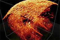
| High-speed imaging with the Bessel beam plane illumination microscope reveals the ever-changing surface of a HeLa cell, with long, thin projections called filopodia continually extending and retracting. |
So at this point, Betzig wondered whether splitting the beam still more might boost the benefits.
Each Bessel beam’s central core is surrounded by concentric rings of weaker light, and he was concerned about how those might interact when the beams began to crowd one another. But when he modeled the interactions among a large field of Bessel beams, his model predicted certain arrangements where the undesirable lobes of light would destroy one another. If he could create those patterns, energy would be spread out, the already low levels of light exposure would drop further, and the pattern would lend itself to enhancing spatial resolution with a technique called structured illumination. “What you get is sort of a triple win,” Betzig says.
Whole New Window
Ten years earlier, before he landed the job at Janelia, Betzig had been in need of a big idea. After several early successes at AT&T’s Bell Labs, where he was the first to image single fluorescent molecules at room temperature instead of in extreme cold, he had become frustrated with academic science. He left Bell Labs and spent the next seven years leading research and development at his father’s machine tool company in Michigan. But that didn’t hold his interest forever. He was ready to return to the lab—but he needed to convince someone to hire him. “I figured it was my last chance to make a scientific career,” he says.
His instinct was to delve into something totally new. But when he learned that biologists could now tag specific molecules inside living cells with fluorescent proteins—a power he’d longed for during his Bell Lab days—he knew it was time to renew his focus on microscopy. He retreated to a cabin in Michigan to immerse himself in theoretical physics and devised a scheme to limit light damage to cells during imaging. The technique he proposed involved illuminating samples with a massive three-dimensional array of light foci. He’d never built that “optical lattice,” as he called it, but now it was time to return to the idea.
“I realized that the ‘magic periods’ [of the theoretical Bessel-beam light field] were the periods that were predicted by my optical-lattice theory 10 years ago,” Betzig says. So he used that theory to map out the light patterns he expected would be optimal for imaging. Once postdoctoral researchers Kai Wang and Bi-Chang Chen figured out how to best generate those patterns, their microscope began producing three-dimensional images of cells with the detail, speed, and low toxicity that they’d hoped for. The microscope is so gentle, Betzig says, that “there are many cells you could look at forever, in 3-D. In a way, it’s pretty much the final solution for that.”
. Image by Kai Wang, Davis Bennett, Misha Ahrens, and Eric Betzig.")
Over the next year, Betzig invited 30 teams of biologists to visit Janelia to work with Chen and fellow postdoc Wesley Legant on the new microscope. Over thousands of hours, they worked out the kinks to better understand the technology’s potential, generating terabytes of breathtaking movies for each group. Those movies were unlike any the scientists had ever seen. The long, thin microtubules that give a cell its form and structural support could be seen rapidly growing, shrinking, and regrowing as cells reshaped themselves and prepared to divide. Individual gene-activating proteins could be followed in three dimensions as they diffused through cells and bound to DNA. Development-regulating growth factors could be monitored for hours as small clumps of cells began to transform themselves into more complex organisms.
“We’ve worked with a new group of biologists every week, and each time it’s a new adventure,” Betzig says. “I’ll never be a biologist, but I get a kick out of the art of it—out of the craziness of the cell. It feels like we have a whole new window on nature that we didn’t have before.”
Now that they have demonstrated a miscellany of applications for the lattice light sheet process, Betzig and his team are eager to continue developing the technology. Their highest priority is integrating a rapid adaptive optics technique that Betzig and Wang developed last year for use in transparent tissues. “We’re going to try to extend the lattice light sheet so we can [image] through the entirety of transparent organisms like the zebrafish or [the roundworm] C. elegans and have all the benefits of the noninvasiveness,” he explains.

Betzig doesn’t want that work to impede researchers’ access to the current lattice light sheet microscope, so his team built a duplicate instrument for Janelia’s new Advanced Imaging Center (see “Microscopes for the Masses”). The center’s director, Teng-Leong Chew, says that months before the lattice light sheet microscope was officially announced, it was already the most in demand of the imaging center’s many cutting-edge technologies. Betzig and his team also helped build two more of the instruments, for labs at Harvard and the University of California, San Francisco. He hopes the lattice light sheet process, which has been licensed to Zeiss, will eventually become standard technology in most labs. But until that time comes, interested researchers have the option of building their own instrument, by following detailed plans from Betzig’s lab.
His team reported on this new microscope in Science on October 24, 2014—just weeks after the Royal Swedish Academy of Sciences had named Betzig a Nobel laureate. In fact, Betzig worried a bit that the recognition for his work on PALM was misguided. “The lattice will be my lasting impact,” he says. “My guess is this will be the high-water mark of my career.”
| This video shows five different stages during the division of a HeLa cell as visualized by the lattice light sheet microscope. Credit: Betzig Lab, HHMI/Janelia Research Campus; Mimori-Kiyosue Lab, RIKEN Center for Developmental Biology; published in Science. |
Though work on the lattice is not yet done, Betzig expects the final optimizations to proceed smoothly. “I’m just dotting i’s and crossing t’s,” he says. “I pretty much know that most of the stuff I’m doing right now is going to work.”
That kind of stability makes Betzig restless. “It’s time to throw the dice again,” he says. “It’s time to try something risky—the kind of stuff [Janelia] is designed to support.”