
Bassett Collection of Stereoscopic Images of Human Anatomy
 David Lee Bassett (1913-1966) was awarded
his MD by Stanford University School of Medicine in 1939. He began
teaching in the Department of Anatomy at Stanford University School
of Medicine in 1939 and later also taught anatomy at the University
of Washington in Seattle. In 1948 Bassett met William B. Gruber,
inventor of the Viewmaster system of stereoscopic imagery. Bassett
and Gruber collaborated on a seventeen-year project of creating
three-dimensional photographic images of human anatomy using
innovations in dissection pioneered by Dr. Bassett.
David Lee Bassett (1913-1966) was awarded
his MD by Stanford University School of Medicine in 1939. He began
teaching in the Department of Anatomy at Stanford University School
of Medicine in 1939 and later also taught anatomy at the University
of Washington in Seattle. In 1948 Bassett met William B. Gruber,
inventor of the Viewmaster system of stereoscopic imagery. Bassett
and Gruber collaborated on a seventeen-year project of creating
three-dimensional photographic images of human anatomy using
innovations in dissection pioneered by Dr. Bassett.
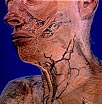
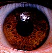
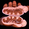
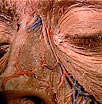 Three artists developed detailed line
drawings based on the photographs: Ruth Ogren, Harriet O’Neill, and
Lorene Segal. 1,547 photographic images and accompanying drawings
were compiled into a 24-volume Stereoscopic Atlas of Human Anatomy,
completed in 1962. The images in this collection are derived from the
Atlas project.
Three artists developed detailed line
drawings based on the photographs: Ruth Ogren, Harriet O’Neill, and
Lorene Segal. 1,547 photographic images and accompanying drawings
were compiled into a 24-volume Stereoscopic Atlas of Human Anatomy,
completed in 1962. The images in this collection are derived from the
Atlas project.
These images are licensed under a Creative Commons Attribution-Noncommercial-Share Alike 3.0 United States License.
Higher resolution images are available for a fee. Contact Drew Bourn, PhD, MLIS.
